Curious about nuclear medicine? It’s a safe way doctors use tiny amounts of radioactive materials to spot problems like heart or lung disease inside your body.
Nuclear medicine is a unique way doctors look inside our bodies using small amounts of radioactive materials. While that might sound scary, these materials are safe and really helpful. They allow doctors to see problems like lung or heart diseases and check how well our organs function.
The tests involve a special camera and, sometimes, a small injection or a pill. Patients usually feel fine during the tests. If you want to learn more about how these fascinating procedures work, keep reading! There’s so much more to discover about the amazing world of nuclear medicine!
Key Takeaway
- Nuclear medicine helps doctors see inside our bodies using small amounts of radioactive materials.
- Procedures like CT scans, VQ scans, and PET scans are important for diagnosing diseases.
- Safety measures are in place to make sure these tests are safe for everyone.
What is Nuclear Medicine?
Nuclear medicine scanning helps doctors see inside the body. A gamma camera, which looks like a big camera on an arm, takes special pictures after patients receive a small amount of radioactive material (about the same radiation as a flight from Sydney to Perth).
Common nuclear medicine scans in Australian hospitals include:
- Bone scans to check sports injuries
- Heart function tests
- Lung examinations
- Thyroid checks
The process takes 30-45 minutes. Patients lie still on a table while the gamma camera moves around them, tracking the radioactive material through their body. The machine doesn’t make much noise, and many patients find it quite relaxing.
After the scan, drinking extra water helps remove the tracer from the body faster. The radiation leaves the body naturally within a day or two. Most Australian hospitals do these scans daily, making them a standard part of medical diagnosis.
How Does It Work?
Nuclear medicine scans use special tracers that glow under medical cameras. The process starts with a tiny injection of technetium-99m, a safe radioactive material that’s no bigger than a teaspoon.
The procedure follows these steps(1):
- A nurse injects the tracer into a vein
- Patient waits 3 hours as the tracer moves through the body
- Patient lies still under a gamma camera for pictures
- Drinking water helps clear the tracer afterward
The tracer acts like a spotlight, showing doctors exactly where to look for problems in bones, hearts, or lungs. While the word “nuclear” might sound worrying, the radiation dose is quite low (about the same as a chest x-ray).
During the waiting period, patients can read or rest quietly. The camera part takes 20-45 minutes, depending on which body part needs scanning. Water intake after the scan speeds up the natural clearing process.
Why is Safety Important?
Nuclear medicine scans make some patients nervous in Melbourne hospitals, but the process is safer than most think. The radiation dose from a thyroid scan sits lower than a Sydney to Perth flight.
Australian hospitals follow strict guidelines set by ARPANSA (Australian Radiation Protection and Nuclear Safety Agency) for these procedures. The doses range from 0.1 to 20 millisieverts, which is quite small in medical terms(2).
Before any scan, staff check important details:
- Pregnancy status
- Breastfeeding status
- Current medications
- Medical history
The thyroid scan uses a tiny iodine-131 pill that helps doctors see how the thyroid works. A gamma camera tracks the iodine as it moves through the gland. Medical staff wear special protective gear and blue dosimeter badges to measure their radiation exposure. For best results, patients should drink plenty of water after the scan to help clear the tracer from their system.
Common Tests in Nuclear Medicine
Sources: Australian Health Journal.
The bustling halls of Royal Melbourne Hospital showcase various medical imaging tools that help doctors see inside the body. Each type of scan works differently, giving specific information about a patient’s health.
Common types of nuclear medicine scans include:
• Bone scans detect fractures and infections using special dye that sticks to damaged areas
• CT (Computed Tomography) creates detailed 3D pictures from X-rays
• PET (Positron Emission Tomography) shows how cells use sugar, helping find unusual growth
• VQ (Ventilation-Perfusion) checks how well lungs move air and blood
CT scans, one of the most common types, take about 10 minutes to complete. The machine produces a radiation dose of 7 millisieverts – equal to about 2 years of natural background radiation. Medical professionals monitor these levels carefully to keep patients safe.
Patients often find these scans quick and painless, though the machines can make loud clicking sounds during the process.
What Are the Benefits?
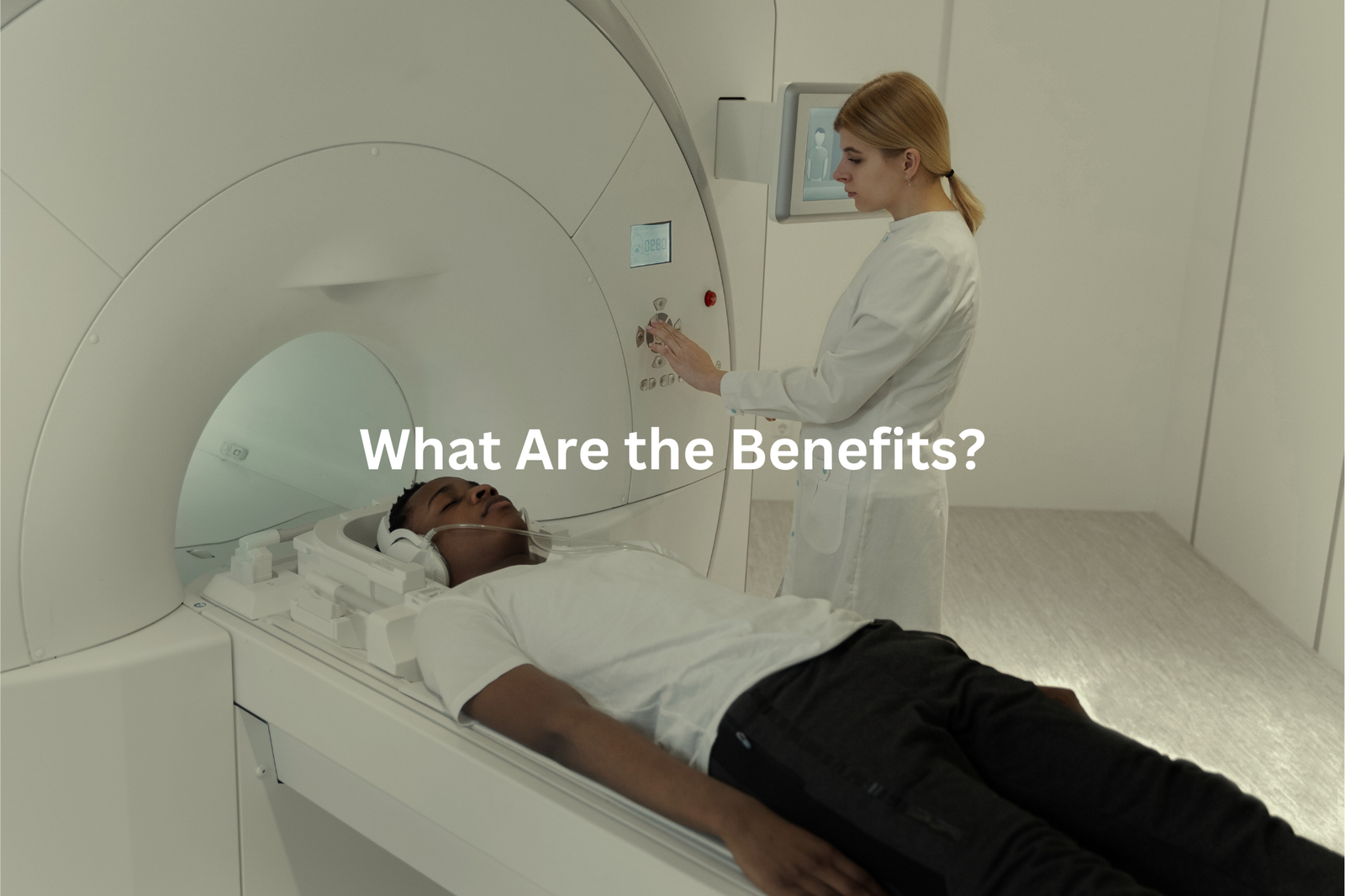
Nuclear medicine scans reveal the inner workings of the human body, creating detailed maps of organs and tissues in real-time. These advanced imaging tests use small amounts of radioactive tracers (roughly equal to the radiation exposure from a Sydney to Perth flight) to track biological processes.
The process starts when a patient receives the tracer through injection or pill form. Special gamma cameras then capture the tracer’s movement, producing clear images of bodily functions.
Common applications include:
- Early cancer detection
- Lung function assessment
- Heart blockage identification
- Bone fracture detection
- Thyroid disorder treatment
The precision of nuclear medicine makes it a valuable diagnostic tool. For thyroid treatments, patients might need to limit close contact with others for several days while the radioactive material naturally breaks down. Modern nuclear scanning equipment can spot issues smaller than a grain of rice, helping medical teams create targeted treatment plans before symptoms worsen.
What About Side Effects?
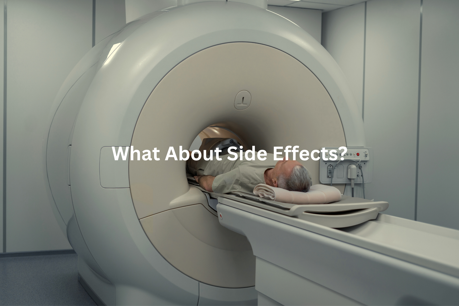
PET scans at Royal North Shore Hospital in Sydney show doctors what’s happening inside a patient’s body. The process uses a small amount of radioactive material (about 7 millisierverts – less than a typical CT scan) to create detailed images.
Most patients don’t feel much different after their scan. Some common reactions might include:
• Mild tiredness, similar to after a busy afternoon
• Slight headache that fades quickly
• Brief metallic taste in the mouth
• Rare allergic reactions (less than 1% of cases)
Medical staff check each patient’s history before the scan, looking at:
• Current medications
• Pregnancy status
• Known allergies
• Medical conditions
The radiation exposure meets Australian safety standards set by ARPANSA. Patients should mention any unusual feelings to their doctor during or after the procedure – medical staff can address concerns straight away. A light meal and plenty of water help most people feel better after the scan.
Future of Nuclear Medicine
Nuclear medicine brings new hope to cancer treatment across Australia(3). At Royal North Shore Hospital in Sydney, doctors now use targeted radiation treatments that work differently from traditional methods.
The star player is Lutetium-177, a tiny radioactive particle just 0.5 millimeters in size. It seeks out prostate cancer cells like a guided missile, leaving healthy tissue alone.
Australian hospitals now offer:
- Radioimmunotherapy that combines radiation with antibodies
- Non-surgical options for skin cancer
- Advanced bone cancer treatments (developed at ANSTO)
“These treatments act like GPS-guided missiles instead of bombs,” explains Dr. Sarah Chen from Royal North Shore Hospital. “The precision keeps getting better.”
The benefits show up clearly in patient care. People spend less time in hospital beds, and many don’t face the harsh side effects common with traditional treatments. While not perfect, these advances mark real progress in Australian cancer care.
FAQ
What is a VQ scan?
A VQ scan, or ventilation-perfusion scan, is a type of imaging test that looks at how well air and blood are flowing through your lungs. It uses small amounts of radioactive gases or liquids to create images that show lung function and blood flow.
What is a lung scan?
A lung scan is a type of medical imaging test that creates pictures of the lungs. It can help identify lung diseases, blood clots, or other issues by looking at how air and blood move through the lungs.
What is a bone scan?
A bone scan is an imaging test that uses small amounts of radioactive material to look at the bones. It can help detect bone injuries, infections, or cancer that may not show up on regular x-rays.
What are PET scans?
PET scans, or positron emission tomography, are imaging tests that use a radioactive substance to look at organ and tissue function. They can help diagnose certain conditions, like cancer, heart disease, and brain disorders.
How do blood flow and air supply imaging tests work?
Blood flow and air supply imaging tests, like VQ scans and lung scans, use radioactive gases or liquids to create images that show how well air and blood are moving through the lungs. This can help identify lung diseases or blood clots.
What is a CT scan?
A CT scan, or computed tomography, is an imaging test that uses X-rays to create detailed, cross-sectional images of the body. It can help diagnose a variety of conditions, from cancer to lung disease.
Can nuclear imaging tests affect breastfeeding?
Nuclear imaging tests, like bone scans or cardiac stress tests, may involve small amounts of radioactive materials. While this is generally safe, it’s important to talk to your doctor if you are breastfeeding to ensure the safety of your baby.
What is a SPECT scan?
SPECT scans, or single-photon emission computed tomography, are a type of nuclear imaging test that uses small amounts of radioactive materials to create 3D images of the body. They can help diagnose conditions like heart disease, brain disorders, and certain types of cancer.
What are the risks of nuclear imaging tests?
Nuclear imaging tests, like PET or SPECT scans, involve small amounts of radiation exposure. While the risks are generally low, it’s important to discuss the potential radiation exposure with your doctor, especially if you are pregnant or have concerns about radiation.
Conclusion
Nuclear medicine is a cool tool for doctors. It lets them look inside our bodies to find health problems early. With careful safety rules (like using protective gear) and constant research, this field is super important in healthcare. If you have questions about a nuclear medicine test, make sure to ask your doctor. They want you to understand what’s happening and feel comfortable. Knowledge is really useful when it comes to taking care of your health!
References
- https://www.prpimaging.com.au/how-nuclear-medicine-works/
- https://www.arpansa.gov.au/sites/default/files/legacy/pubs/rps/rps14_2.pdf
- https://www.minister.industry.gov.au/ministers/husic/media-releases/securing-future-critical-nuclear-medicines

