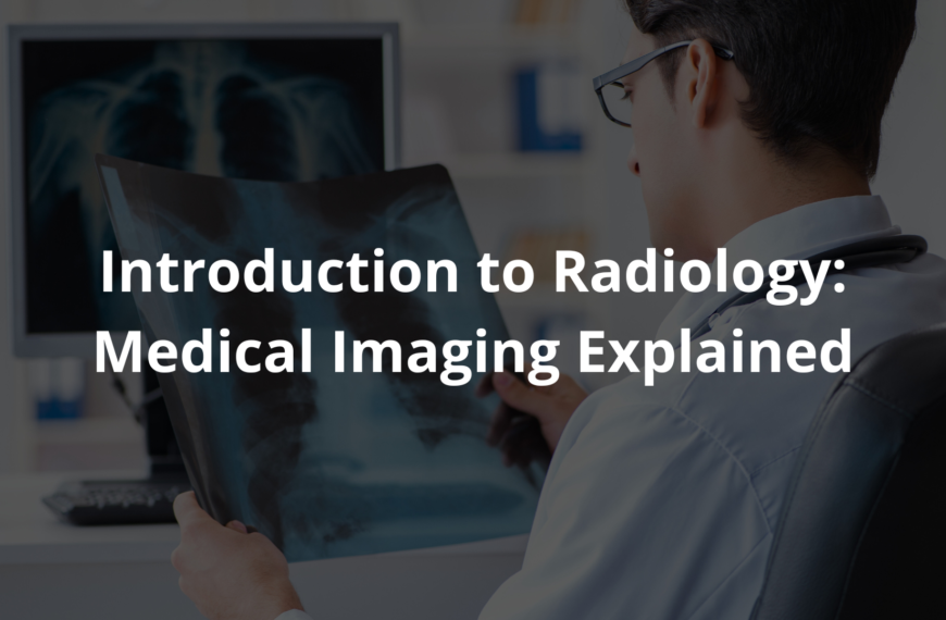
Discover how radiology advancements in Australia are helping doctors detect problems faster, saving lives with clearer, more precise imaging.
Radiology advancements are like using special glasses to look inside our bodies. New technology provides clearer images, helping doctors find problems quicker. This is super important because discovering health issues early can save lives. For example, 3D imaging allows doctors to see details they couldn’t before, improving diagnosis accuracy.
Plus, AI tools can analyse scans faster, so patients get results without waiting long. These changes help doctors do their jobs more efficiently and provide better care for everyone. There’s so much more to learn about how radiology is shaping healthcare today. Keep reading to find out more!
Key Takeaway
- New imaging systems help doctors see inside the body clearly.
- AI can find problems in medical images quicker than the human eye.
- Early detection of diseases like cancer can lead to better patient care.
Digital Imaging Systems: A New Way to See Inside
I’ve been noticing something pretty amazing happening in medical imaging, especially here in Australia. It’s like science fiction coming to life. With digital technology, doctors can now see inside the human body in ways that were impossible just a few years ago. It’s fast, detailed, and honestly, kind of mind-blowing.
Radiologists (they’re the doctors who read these images) use tools like X-rays, MRIs, and CT scans to figure out what’s going on inside us. Each tool works differently—X-rays are great for bones, MRIs are better for soft tissues like your brain, and CT scans give a 3D view of everything. It’s like having a toolbox where each tool does something special.
The real game-changer, though, is how quickly these images can be shared. A doctor in Sydney can send a scan to a specialist in Melbourne in seconds. And with artificial intelligence (AI) lending a hand, they can catch tiny problems that might’ve been missed before. For example, AI can help spot early signs of cancer, giving patients a better chance at treatment.
But safety is a big deal too. Radiologists are trained to keep radiation levels as low as possible (it’s called ALARA—As Low As Reasonably Achievable). They follow strict rules to ensure patients are safe during scans.
So, if you ever need an X-ray or MRI, don’t be afraid to ask questions. These tools are here to help, and you’re the most important part of the whole process.
Artificial Intelligence: The Brain Behind the Images
Source: Sky News Australia.
AI is kind of like having a super-smart helper for radiologists. Picture a teacher in a classroom who’s great at catching mistakes in maths problems—that’s what AI does with medical images. When doctors review CT scans (which are detailed pictures of the inside of the body), AI steps in to help spot things that might be easy to miss.
For example, if a radiologist is checking a scan for signs of lung cancer, AI can highlight unusual areas that might need extra attention. This can make a big difference for patients. It means quicker results and fewer errors. It’s like having an extra pair of sharp eyes that never get tired.
AI algorithms (the step-by-step instructions that help AI work) are improving all the time. As they learn and adapt, they can assist doctors even more. This teamwork between AI and radiologists is making diagnosing illnesses faster, safer, and more accurate.
Advanced Imaging Techniques: Different Ways to See
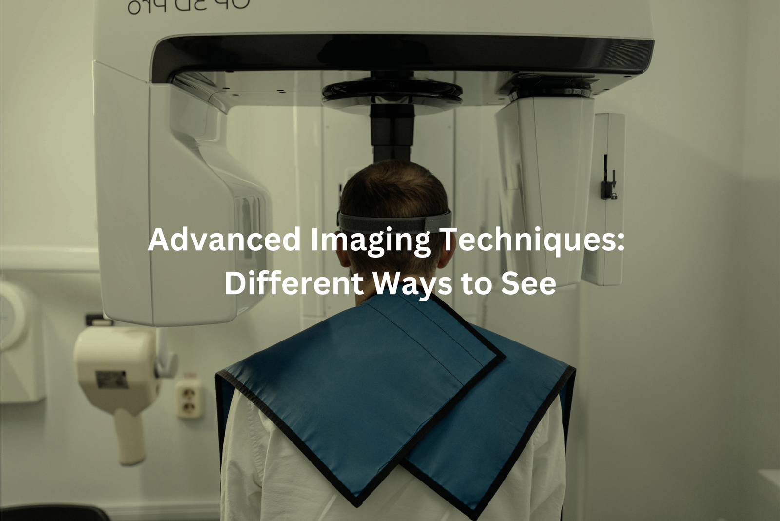
Radiologists have some pretty incredible tools to peek inside our bodies, and each one has a specific job. It’s like having a toolbox, but for medicine. Here are the main ones they use:
- X-rays: These are quick and great for looking at bones. If you’ve ever broken an arm or leg, chances are you’ve had an X-ray. They can also spot infections or other bone problems.
- MRI (Magnetic Resonance Imaging): This one’s special because it doesn’t use any radiation. It’s brilliant for checking soft tissues, like your brain, muscles, or even your heart.
- CT (Computed Tomography) Scans: CT scans are like super-detailed X-rays. They take lots of pictures from different angles to create a 3D image. Doctors often use them to find things like tumors or internal injuries.
- Ultrasound: This tool uses sound waves to make pictures. It’s famous for showing babies during pregnancy, but it’s also handy for looking at organs like the liver or kidneys.
Each tool has its strengths, and together they help doctors figure out what’s going on inside. If you’re ever curious, ask your doctor about the imaging they’re using—it’s fascinating stuff.ent care. It’s like a symphony, where each instrument adds to the beautiful music of healthcare.
Safety Measures: Keeping Patients Safe
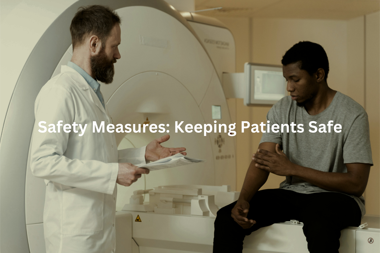
Patient safety in radiology is a big deal(1). It’s like having a team of heroes working together to keep everyone safe. Radiologists and doctors use special equipment, like lead aprons and shields, to protect themselves and their patients from too much radiation. Think of it as wearing armour that blocks out danger.
Before ordering an X-ray or scan, doctors carefully decide if it’s really needed. They weigh up the benefits (like spotting an illness early) against any risks. This way, patients only get the imaging they truly need. It’s all about being smart and cautious.
The machines used for imaging, like X-ray or CT scanners, are checked regularly. It’s a bit like tuning up a car to make sure it runs smoothly. Radiologists make sure everything’s working properly so patients can feel safe.
If you ever need an X-ray or scan, know that the team is focused on your safety. Trust the process, ask questions if you’re unsure, and let the experts take care of the rest.
Importance of Early Detection: Catching Problems Early
You know, catching health problems early can really make a difference. It’s like spotting a tiny leak in a boat—if you fix it right away, you might avoid sinking. For things like lung cancer or breast cancer, early detection can mean the difference between a tough battle and a much easier one.
Doctors have some pretty amazing tools for this, like X-rays, MRIs, and ultrasounds. These machines can see things inside your body that you wouldn’t even know were there. For example, if a doctor finds a tumour when it’s still small (maybe just a few millimetres), there’s often a better chance of treating it successfully. That’s a big deal.
So, it’s really important to talk to your doctor about regular screenings. Think of it like checking your car’s engine before a long trip—it’s just smart. Regular check-ups and tests could save your life. Don’t wait for something to feel wrong.
Continuous Education: Keeping Up with the Times
Radiologists don’t just stop learning once they’ve got their medical degree. It’s like they’ve signed up for school forever, but they actually seem to like it. They’re always attending workshops and training sessions to keep up with the latest technology and techniques(2).
Sometimes, they’ll travel long distances—hours or even days—to go to conferences. These events are packed with research papers (usually with really complicated titles) and presentations about the newest discoveries. This ongoing education is what helps radiologists provide the best care for their patients. When new machines or methods come along, they’ve got to know how to use them properly to help people heal.
Radiologists also work together, sharing what they’ve learned with each other. They meet in groups, almost like a big study club, to exchange ideas and improve as a team. This teamwork means better care for patients, which is the whole point, really. So, if you see a doctor chatting with another, they’re probably swapping tips to help someone. Keep learning, keep sharing—that’s how they stay sharp.
FAQ
How can low-dose ray tube technology improve radiology?
Low-dose ray tube technology in modern CT scanners and other imaging systems can significantly reduce the radiation dose patients receive during medical scans, without compromising image quality. This helps minimise the health risks associated with repeated exposure to ionising radiation, leading to safer and more comfortable patient experiences.
What are the benefits of real-time imaging in radiology?
Real-time imaging techniques, such as fluoroscopy, allow radiologists to observe anatomical structures and physiological processes in near real-time. This enables more accurate diagnoses, better guidance during interventional procedures, and faster decision-making, ultimately leading to improved patient care and outcomes.
How do wide-range flat-panel detectors enhance digital radiography?
Flat-panel detectors used in digital radiography (DR) systems offer a wide dynamic range, allowing them to capture high-quality images across a broad spectrum of radiation doses. This flexibility improves image quality and reduces the need for repeat scans, benefiting both patients and healthcare providers.
How can AI systems help with medical image data analysis?
Radiology AI systems are trained to rapidly analyse large volumes of imaging data, detecting subtle patterns and abnormalities that may be missed by the human eye. This AI-powered image analysis can assist radiologists in making more accurate diagnoses, streamlining workflows, and ultimately delivering better patient care.
What role does dual-energy CT play in modern radiology?
Dual-energy CT scanners use two different X-ray energies to capture images, enabling the differentiation of materials like bone, soft tissue, and contrast agents. This advanced imaging technique provides valuable information about the composition of anatomical structures, supporting more precise diagnoses and treatment planning in fields like oncology and cardiology.
How do third-party cloud storage solutions benefit radiology practices?
Cloud-based storage and data management platforms allow radiology practices to securely store and access medical images, reports, and patient data from anywhere. This improves workflow efficiency, collaboration between healthcare providers, and access to patient information, ultimately enhancing patient care and outcomes.
What are the advantages of open access to medical imaging data?
Sharing medical imaging data through open-access platforms can foster collaboration and innovation in the radiology field. Researchers and clinicians can access a wider range of data to develop new imaging techniques, improve computer-aided diagnosis, and advance our understanding of various medical conditions.
How can radiology AI technologies help improve patient care?
Radiology AI systems can assist in automating time-consuming tasks, such as image segmentation and lesion detection, freeing up radiologists to focus more on patient interaction and clinical decision-making. Additionally, AI-powered tools can provide rapid, standardised interpretations of imaging exams, helping to expedite diagnoses and treatment plans.
Conclusion
Radiology is changing fast, and it’s helping doctors care for us better. New digital systems, smart technology like AI, and better imaging tools help them look inside our bodies clearly. These advancements can find health problems early, make procedures safer, and lead to healthier lives. It’s pretty fascinating to think about how these tools will keep getting better. So, when you hear about radiology, remember how it’s making health care smarter and safer for everyone every day!
References
- https://insightsimaging.springeropen.com/articles/10.1186/s13244-019-0721-y
- https://www.sciencedirect.com/science/article/abs/pii/S1546144004003734