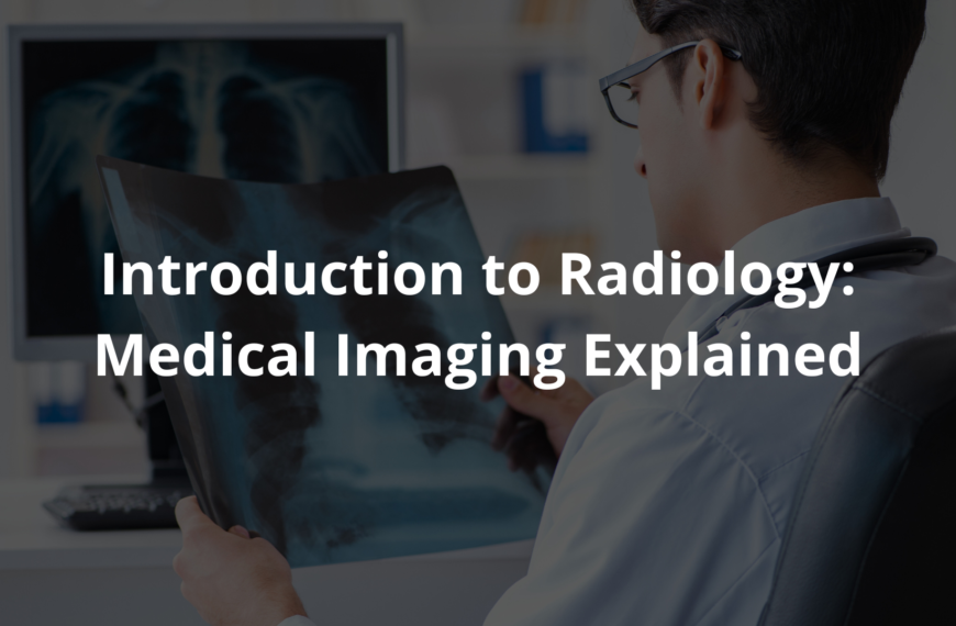
Uncover the exciting journey of radiology from X-ray discovery to modern imaging techniques and how they help doctors.
Radiology history is like a fascinating puzzle, where each piece fits together to reveal the bigger picture of how we see inside the human body. It all began in 1895 with a curious man named Wilhelm Conrad Röntgen. While experimenting with cathode rays, he stumbled upon something extraordinary: X-rays! These rays allowed doctors to look inside patients without needing to cut them open. So, let’s take a closer look at this captivating story of radiology!
Key Takeaway
- X-rays were discovered in 1895 by Wilhelm Conrad Röntgen, leading to a whole new way of seeing inside the body.
- Radiology evolved from simple X-rays to advanced techniques like CT scans and MRIs.
- Many brilliant pioneers helped shape the field of radiology into what it is today.
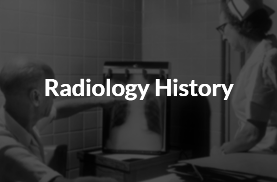
History of X-rays
In 1895, a curious German physicist named Wilhelm Conrad Röntgen was playing with cathode rays. Imagine Röntgen in his lab, surrounded by glass tubes and strange equipment, when suddenly a fluorescent screen began to glow. It was like magic! He hadn’t even pointed the cathode rays directly at the screen.
This unexpected light led him to wonder if there was a new type of radiation. Something unexplained was happening. He decided to call these mysterious rays “X-rays,” with “X” symbolising the unknown.
By January 1896, Röntgen’s discovery was making waves. Doctors, eager to help their patients, began using these X-rays to see inside the human body. They could now diagnose broken bones and locate foreign objects without performing surgery. It was revolutionary! Imagine being able to tell if a bone was broken just by looking at a picture instead of cutting someone open. Röntgen’s invention meant that patient care was changing.
Doctors were thrilled. With X-rays, they could quickly diagnose conditions like fractures(1). It saved time and made procedures safer. Imagine a world where you could walk into a hospital, and with just a quick image, doctors could see what was wrong inside your body. It made everything feel simpler and more efficient. There was a new tool in the medical toolbox, and it was opening doors to new possibilities.
Invention of Radiology
After Wilhelm Röntgen’s remarkable discovery, radiology began to take shape as a medical field. It was like watching a new plant sprout and grow. The very first medical X-ray in the United States was taken by a doctor named Gilman Frost. Picture him at Dartmouth College, using X-rays to look at a broken wrist.
This was a pivotal moment because it demonstrated how valuable X-rays could be in medicine. Suddenly, doctors had a way to see inside the body without invasive procedures.
With this new ability, doctors quickly realised they could diagnose patients in a much more efficient way. Instead of guessing, they could look right at the problem. This made the job of being a doctor a bit easier, I think. Patients no longer had to worry about the risks that come with surgery just to find out what was wrong. It was a fantastic leap forward in medical technology.
Radiology continued to evolve as more doctors embraced the technology. X-rays opened the door for further advancements. Who would have guessed that such a simple discovery would lead to a whole field of medicine? The potential was enormous. From that point on, doctors began to explore new ways to create images of the human body. They were like explorers charting unknown territory.
As radiology grew, it became an essential part of healthcare. It helped doctors understand what was happening inside their patients. It transformed the way healthcare was delivered. Today, radiology plays a crucial role in diagnosing and treating conditions. The journey from Röntgen’s first X-ray to the established field of radiology is a fascinating story of discovery, innovation, and the desire to help others.
Evolution of Imaging
Radiology didn’t stay the same; it grew and changed over the years. Here are some major advancements:
- Fluoroscopy: Thomas Edison played a key role in developing this technology, which allowed real-time imaging. Imagine watching a video of your organs moving!
- CT Scans: Introduced in the 1970s, CT (Computed Tomography) scans provided doctors with cross-sectional images using X-rays. This gave a new perspective on the human body, like slicing a loaf of bread to see what’s inside.
- MRI: In the 1980s, magnetic resonance imaging (MRI) became widely used. It provides detailed images of soft tissues without using radiation, making it safer for patients.
Early Diagnostic Techniques
In the early days of radiology, X-rays were like a new pair of glasses for doctors. They helped doctors see inside the human body in a way that was never possible before. Picture a doctor in a small hospital, looking at a plate with an X-ray image of a broken bone. It must’ve felt like magic to suddenly have this new tool at their disposal.
X-rays became a quick way to diagnose issues. They were mostly used to see bones and locate foreign objects that shouldn’t be there, like the classic story of a child swallowing a coin.
Doctors quickly recognised the value of X-rays. They could locate gunshot wounds in emergency situations, which was crucial for saving lives. Imagine a soldier coming into a field hospital with a bullet lodged in their side. Instead of guessing where the bullet was, the doctor could take an X-ray and see exactly where to operate. This was revolutionary and changed the way injuries were treated.
By June 1896, it was even discovered that X-rays could treat skin problems. This opened a whole new world of possibilities, showing that X-rays weren’t just for diagnosis but could also aid in healing.
I think it’s fascinating how quickly medicine adapted to this new technology. Doctors were learning to trust X-rays for things like diagnosing pneumonia, which was a common illness at the time. They could now see the lungs and identify what was going wrong. This was a big step forward, and it made doctors feel like they had superpowers. As they explored the uses of X-rays, doctors realised they weren’t just looking at bones anymore.
Radiology Advancements

As time went on, radiology continued to improve with new technologies:
- Ultrasound: This uses sound waves to create images of soft tissues. It’s often used in pregnancies to see babies before they are born.
- Nuclear Medicine: This technique uses tiny amounts of radioactive materials to help doctors see how organs are working. It can even treat certain illnesses.
- Digital Radiography: Instead of using film, digital imaging systems are now common, allowing for quicker results and less radiation exposure.
Radiology Pioneers
Several key figures made significant contributions to radiology:
- Wilhelm Conrad Röntgen: The father of X-rays, he received the first Nobel Prize in Physics in 1901.
- Thomas Edison: He didn’t just invent light bulbs; he also helped improve fluoroscopy, which allowed doctors to see live images.
- Sir Godfrey Hounsfield: He developed the first CT scanner, revolutionising how doctors could view internal structures.
Radiology Timeline
Here’s a quick timeline to highlight the key moments in radiology history:
- 1895: Röntgen discovers X-rays.
- 1896: The first clinical use of X-rays in medicine.
- 1918: Film is introduced for radiography.
- 1972: The first CT scan is performed.
- 1980s: MRI technology becomes widely used.
Imaging Milestones
Radiology has seen many exciting milestones:
- The introduction of digital imaging techniques, which improved efficiency.
- The development of hybrid imaging systems, combining different methods for better results.
- Advances in software that help doctors interpret images more accurately.
Modern Radiology
Today, radiology is an essential part of medicine, using advanced techniques and technologies:
- Digital Imaging and Communication in Medicine (DICOM): This standard helps store and share medical images easily.
- Artificial Intelligence: Computers now help analyse images and assist in diagnosing conditions, making the process faster and more efficient.
- Minimally Invasive Procedures: With imaging techniques, doctors can perform surgeries with smaller cuts, leading to quicker recoveries for patients.
History of MRI
The history of MRI is a fascinating journey of innovation and discovery. In the late 20th century, Dr Raymond Damadian proposed using magnetic fields for imaging, paving the way for a new era in medicine. The first human MRI scan took place in 1977, and it was a game-changer(2).
MRI technology allows doctors to look at soft tissues like the brain and joints without using harmful radiation. This is crucial for diagnosing conditions that X-rays might miss. Imagine a patient with a mysterious illness lying inside a large machine, surrounded by a powerful magnet. The machine hums and clicks, capturing detailed images of their insides.
MRI is fantastic because it provides detailed views of the body, helping doctors make informed decisions. This non-invasive tool has saved countless lives and continues to be a vital part of modern medicine. The journey from the first scan to today’s advanced imaging techniques is truly remarkable.
Conclusion
In wrapping up the story of radiology history, we’ve seen how it all started with Röntgen’s groundbreaking discovery of X-rays. This led to the evolution of radiology, with many remarkable advancements and pioneers shaping the field.
Today, radiology plays a crucial role in patient care, helping doctors diagnose and treat various conditions more effectively. So, the next time you hear about an X-ray or an MRI, remember the incredible journey that brought us to this point!
FAQ
How did Wilhelm Conrad Röntgen revolutionise medical imaging with his discovery of X-rays?
In 1895, Wilhelm Conrad Röntgen discovered X-rays while experimenting with cathode ray tubes. He noticed that a ray beam could pass through soft tissue but not bones, creating ray images on photographic plates. This breakthrough earned him the first Nobel Prize in Physics and launched the history of radiology.
How did CT scanning evolve from basic ray imaging to computed tomography?
Sir Godfrey Hounsfield and Allan Cormack developed CT scanning in the 1970s. Their work with CT scanners transformed medical imaging by creating images of the human body in cross-sections. The CT imaging technology uses ionising radiation to produce detailed images, earning them the Nobel Prize in Physiology or Medicine.
What role do diagnostic imaging modalities play in modern medicine?
Today’s imaging techniques include projectional radiography, magnetic resonance imaging, computed tomography, and ultrasound imaging. Each imaging modality serves different purposes in diagnostic radiology. MRI scans use nuclear magnetic resonance for soft tissue imaging, while PET CT scans help detect diseases using positron emission tomography.
How has digital imaging changed radiology departments?
Digital radiography and digital imaging replaced photographic film in radiology departments, improving image quality and patient care. Real-time imaging allows for minimally invasive procedures in interventional radiology. The shift to artificial intelligence in imaging interpretation marks the latest advancement in diagnostic imaging.
What training programs prepare doctors for radiology careers?
Radiology residency programs at university schools of medicine combine medical education with hands-on training. The Royal Australian and New Zealand College of Radiologists oversees training standards. Residents learn radiation safety, nuclear medicine, paediatric radiology, and emergency radiology. Clinical radiology training includes breast imaging, bone densitometry, and radiation protection.
How has radiation protection evolved in medical procedures?
Medical physics experts developed radiation protection standards to minimise the effects of radiation exposure. Understanding electromagnetic radiation and types of radiation led to safer protocols. Contrast agents and dual energy techniques help optimise imaging while reducing radiation doses during medical procedures.
References
- https://en.wikipedia.org/wiki/X-ray
- https://columbiasurgery.org/news/2015/09/17/history-medicine-dr-roentgen-s-accidental-x-rays