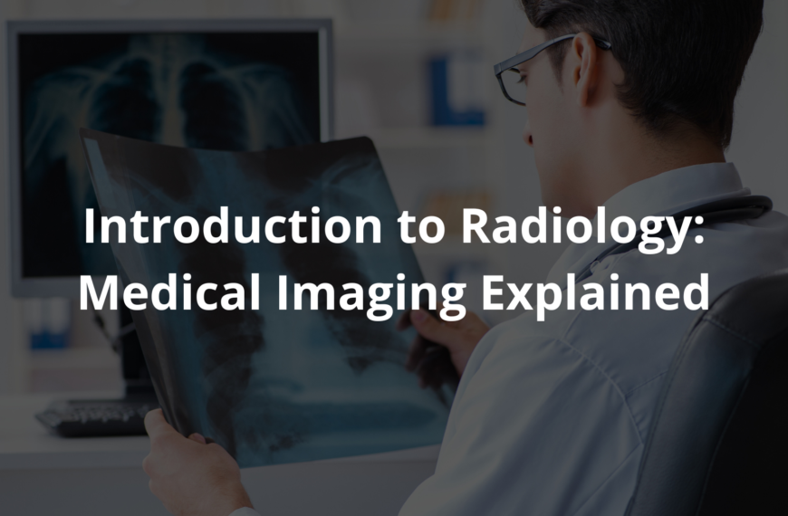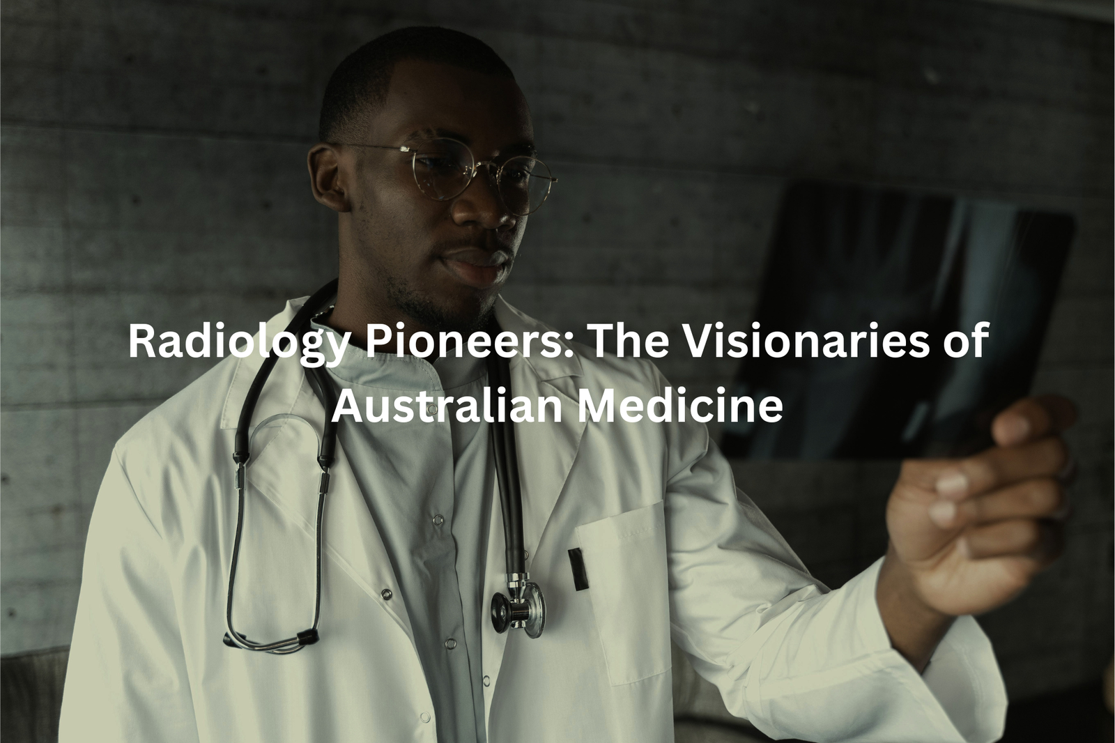
Meet the radiology pioneers who changed healthcare forever, bringing X-rays and imaging to life in Australia for better patient care.
Radiology pioneers changed the game for doctors and patients, kinda like superheroes. Thanks to Wilhelm Conrad Röntgen, who discovered X-rays in 1895, we can now peek inside our bodies without surgery. In Australia, many brave innovators joined this journey, bringing medical imaging to life in hospitals and clinics everywhere.
They developed new techniques and machines that help detect illnesses earlier and treat them better. Some famous names include Dr. John Flynn, who used X-rays in remote areas, and Dr. John Corboy, who advanced imaging technology. If you’re as curious as I am, keep reading to uncover more about these legends!
Key Takeaway
- Radiology pioneers helped create X-ray technology that changed patient care.
- They worked hard to make MRI and CT scans safer and better.
- Ongoing education is important for radiologists to keep improving.
Early Adoption of X-Ray Technology
When Wilhelm Röntgen discovered X-rays in 1895, it was like finding a secret key to look inside the human body. In Australia, doctors quickly saw how this could change medicine. They no longer had to rely only on guesses or painful surgeries to figure out what was wrong. X-rays let them see inside without cutting, which meant faster diagnoses and better treatments.
By 1949, the Royal Australian and New Zealand College of Radiologists was set up to train people in using X-rays and other imaging tools. Over time, Australian scientists helped develop even more advanced machines, like MRI and CT scanners. These tools give clearer pictures with less radiation, making them safer for patients.
Today, radiologists in Australia are using Artificial Intelligence to improve how they read scans. They’re also careful to minimise radiation exposure, keeping safety a top priority. So, next time you get an X-ray, remember—it’s part of a long history of innovation, and it’s helping doctors take better care of you.
The Royal Australian and New Zealand College of Radiologists
Source: Choosing Wisely Australia.
I remember hearing about the Royal Australian and New Zealand College of Radiologists (RANZCR) during a lecture on medical specializations. It’s like a big training hub for radiologists—doctors who focus on medical imaging. They’ve been around since 1949, which means they’ve had decades to perfect how they teach and guide radiologists. Pretty cool, right?
Radiologists don’t just learn how to use machines like X-rays, MRIs, and CT scanners. They also study how to read the images these machines produce. It’s not as simple as it sounds. Those images can show everything from broken bones to hidden diseases. Training to become a radiologist can take over 10 years (if you count medical school and all the extra training). That’s a lot of studying.
The college also makes sure radiologists stay updated on new techniques and technologies. They even focus on safety—like how much radiation is safe for patients. This is a big deal because it helps protect people during tests. So, next time you’re getting an X-ray or scan, you can feel a bit better knowing these doctors are highly trained and following strict rules.
Contributions to MRI Development
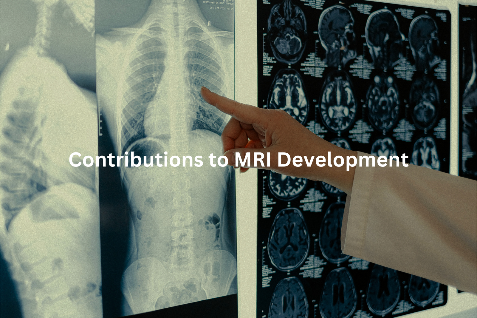
Have you ever wondered how MRI machines work? They’re kind of like X-rays, but way more advanced. Instead of just showing bones, MRIs are brilliant at capturing detailed images of soft tissues—like muscles, organs, and even the brain. That’s why doctors use them to spot things like tumours or injuries that other scans might miss.
In Australia, researchers have been working hard to make MRI technology even better. They’ve figured out ways to make the images sharper and the scans faster. (Imagine taking a blurry photo and tweaking the camera settings until it’s crystal clear—that’s sort of what they’re doing with MRIs.) Faster scans mean less time lying still in that noisy tube, which is a win for patients.
Here’s why this matters: If a doctor sees a tiny tumour on an MRI, they can act quickly. Early treatment often means better outcomes. These improvements could save lives, plain and simple. So, next time you hear about medical research, think about how these small tweaks—like better MRIs—can make a big difference. It’s exciting to think about what might come next.
Advancements in CT Scanning
When I think about CT scans, I can’t help but feel a bit in awe. They’re like these clever machines that can peek inside our bodies without even touching us. It’s kind of like taking hundreds of photos from every angle and then piecing them together into a full picture. That’s how doctors can see what’s going on inside, whether it’s a broken bone or something deeper.
Australia has been a leader in making CT scans safer. Researchers here have worked out ways to get clearer images while using less radiation (which is super important because too much radiation can be harmful). I remember when my uncle had to get a CT scan after a car accident. The doctors were able to figure out what was wrong quickly, and he didn’t have to worry about being exposed to too much radiation.
CT scans are now a trusted tool for doctors. They’re fast, safe, and give life-saving information. If you ever need one, just know that the technology is improving all the time, thanks to smart people working hard behind the scenes. My advice? If you’re ever nervous about it, ask questions. Doctors are always happy to explain how it works.
Artificial Intelligence Integration
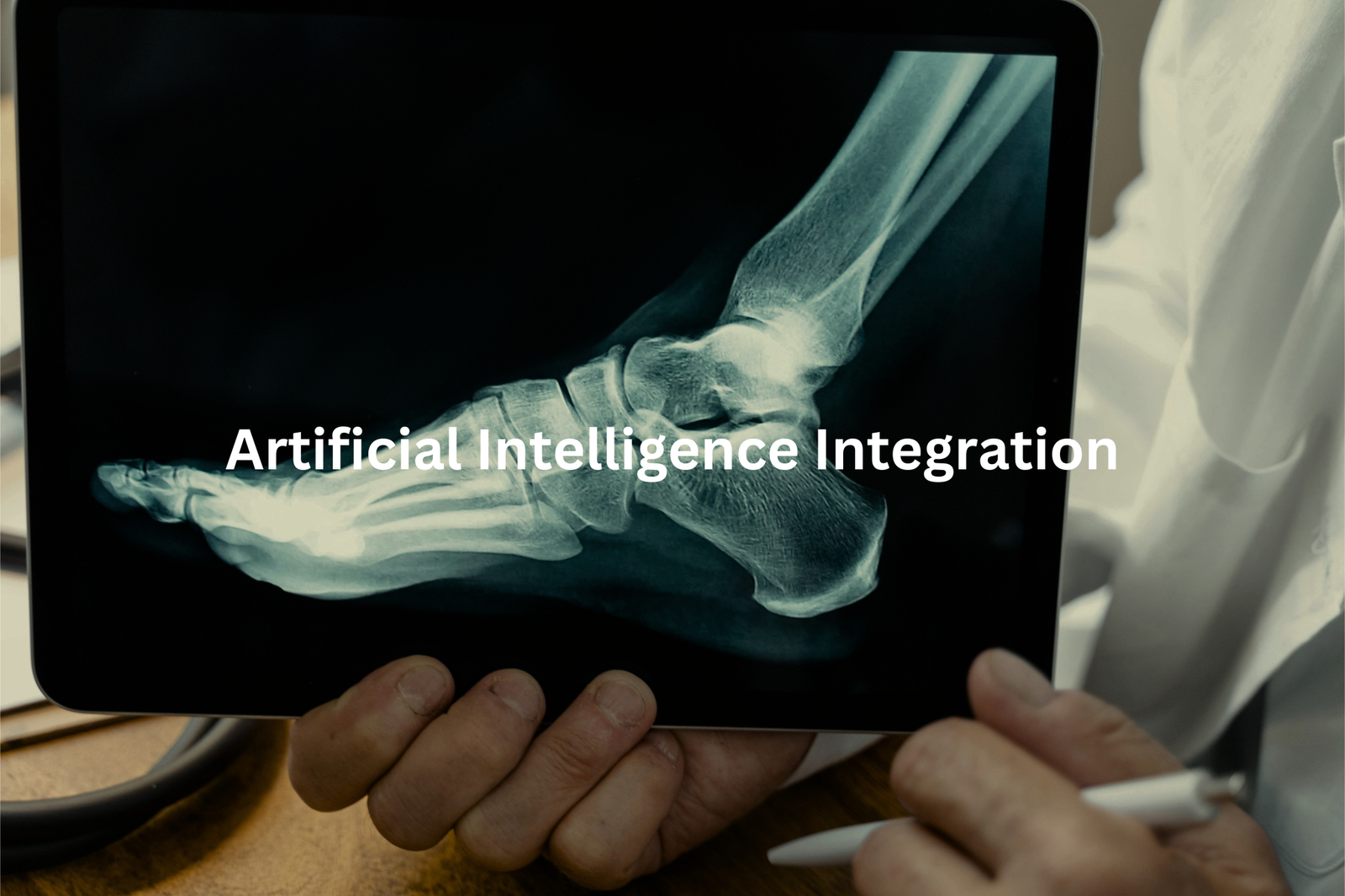
Did you ever think machines could lend a hand to doctors? Well, they’re doing just that in Australia! Artificial intelligence, or AI, is like a clever assistant for radiologists. It examines medical images—like X-rays, CT scans, and MRIs—and spots things that might be wrong, such as tumours or fractures. Think of it as having a super-focused helper that never gets tired or distracted.
Here’s the thing: AI doesn’t replace doctors. Instead, it works alongside them. The AI scans the images and flags anything unusual, like a shadow that could mean trouble. Then the radiologist steps in to review and decide what’s next. It’s teamwork, really—human expertise and machine precision.
This technology can save heaps of time. For example, instead of waiting days for results, patients might get answers much sooner. That’s a big deal, especially if someone’s worried about their health.
In hospitals, AI is already making a difference. Doctors can spend more time with patients, and treatment can start faster. It’s not just about speed, though; it’s about improving care. So, while AI might sound futuristic, it’s already here, quietly transforming healthcare in ways that matter.
Radiation Safety Advocacy
Radiation might sound like a big, complicated word, but it’s actually just energy moving through space. In radiology (that’s the part of medicine where doctors use images like X-rays or CT scans to see inside your body), radiation is super important(1). But here’s the thing—too much of it isn’t good for you. That’s why doctors follow something called the ALARA principle. It stands for “As Low As Reasonably Achievable.” Basically, it means they only use the smallest amount of radiation needed to get the job done.
Think of it like adjusting the brightness on a torch. You don’t need it on full blast if a dim light will do. For example, during a CT scan, doctors carefully plan the settings so they get clear images without overdoing the radiation. It’s a balancing act, and it’s all about keeping you safe.
This approach didn’t just happen overnight. The early scientists and doctors who worked with radiation realised how important it was to protect patients. They set the groundwork for how we use imaging today. So next time you or someone you know needs an X-ray or scan, remember—there’s a whole system in place to make sure it’s done safely. Trust the process, and don’t be afraid to ask questions if you’re curious!
Focus on Early Detection
One thing I’ve noticed about radiology is how it’s like a quiet superhero in healthcare. It helps catch problems early, sometimes even before you feel sick. In Australia, radiologists are big on early detection. They’re kind of like those mates who remind you to put sunscreen on before heading to the beach—not pushy, just looking out for you. They encourage regular screenings, especially for things like breast cancer, because finding something early can make all the difference.
Think about it like spotting a tiny crack in your car’s windshield. If you fix it quickly, it won’t spread and ruin the whole thing. That’s what early detection does for your health—it stops little problems from becoming big ones. And honestly, that’s a relief for anyone who’s worried about their health.
Radiologists also work hard to spread awareness(2). They explain why regular check-ups matter, so more people understand how screenings can save lives. It’s not just about fixing problems; it’s about preventing them. So, if you’ve been putting off a scan or check-up, maybe it’s time to book one. It’s a small step that could keep you healthy longer.
Ongoing Education and Research
Radiology, I reckon, is like piecing together a giant, never-finished jigsaw puzzle. There’s always something new to figure out, which is why education is so important for radiologists. These doctors don’t just stop learning after med school. They’re always sharpening their skills, attending workshops and conferences (some big ones happen in places like Sydney or Melbourne) to stay on top of the latest tools and techniques.
Think of it like athletes training for a big game—they’ve got to keep pushing themselves to be better. Radiologists do the same, but with their minds. Say a new imaging method comes out, one that can detect diseases earlier than before. That’s a game-changer for patients because it means quicker treatment and better outcomes.
By keeping up with the latest advancements, Aussie radiologists make sure they’re giving patients the best care possible. So next time you’re getting a scan, you can feel confident knowing your radiologist is probably well-trained and up-to-date. Lifelong learning—it’s just part of the job.
Interdisciplinary Collaboration
When I think about radiology, I picture a team of people solving a mystery together. It’s not just one person looking at scans. It’s a group effort, where everyone has a role to play. Like putting together a puzzle, each person brings their piece to complete the picture.
Radiologists often work closely with other medical professionals. For example, if someone has a car accident and hurts their chest, a radiologist might study a CT scan to check for injuries. Then, they’d talk with a trauma surgeon about what they see. Together, they’d decide the best treatment to help the patient heal. It’s not just about looking at pictures—it’s about making decisions as a team.
In Australia, radiologists know that sharing knowledge is key. By working together, they can combine their skills and experience to give patients the best care possible. Teamwork in radiology isn’t just helpful—it’s essential.
Global Influence
Standing in a hospital hallway, you can feel the buzz of activity around Australian radiologists. They’re not just taking X-rays or reading scans; they’re shaping how health care works, not just here in Australia but across the world. It’s kind of amazing when you think about it—how their work reaches so far.
A lot of these radiologists publish their research in major international medical journals (the kind doctors everywhere read)(3). They team up with experts from other countries, swapping ideas and learning new techniques. This sharing of knowledge often leads to better ways of diagnosing and treating patients.
What’s even cooler is that Australian radiologists help set the standard for care. Their guidelines are used in places like Europe, Asia, and beyond. So, next time you’re in a hospital, think about this: the scan you’re getting or the treatment plan your doctor suggests might’ve been influenced by someone working in a lab or clinic thousands of kilometers away. Health care’s a team effort, and radiologists are a big part of that team.
FAQ
What is the history of the X-ray tube?
The X-ray tube was invented by physicist Wilhelm Roentgen in 1895. He discovered that by running an electric current through a vacuum-sealed glass tube, it produced a new type of invisible ray that could pass through human flesh, revealing the bones inside. This groundbreaking discovery revolutionised the field of medical imaging and led to the development of the modern X-ray tube used in radiology today.
Who were the pioneers in the field of radiology?
The pioneers of radiology include scientists like Wilhelm Roentgen, the discoverer of X-rays, as well as Marie Curie, who conducted extensive research on radioactivity. Other pioneers include Dr. Joseph Lister, who used X-rays for patient care, and Dr. Edward Jenner, who developed the first vaccine. These individuals made crucial contributions that paved the way for the advancements in the field of radiology we see today.
How did radiology pioneers help during wartime?
During times of war, radiology pioneers played a crucial role in providing medical care. They used their expertise to set up field hospitals and deploy mobile X-ray equipment to treat injured soldiers. For example, during World War I, Marie Curie and her daughter Irene worked tirelessly to bring X-ray technology to the front lines, earning them the nickname “The Radiological Sisters.” Their efforts helped save countless lives by improving battlefield medical care.
What is the significance of the history of radiology?
The history of radiology is significant because it showcases the remarkable achievements of pioneers who pushed the boundaries of science and technology to improve patient care. From the groundbreaking discovery of X-rays by Wilhelm Roentgen to the advancements in magnetic resonance imaging (MRI), the field of radiology has consistently evolved to provide better diagnostic tools and treatments. Understanding this rich history helps us appreciate the dedicated work of radiology pioneers and inspires us to continue advancing the field for the benefit of humanity.
How has radiology technology evolved over time?
Radiology technology has undergone remarkable advancements since the discovery of X-rays. From the early days of photographic plates and cathode ray tubes, the field has progressed to include sophisticated imaging techniques like computed tomography (CT) scans, positron emission tomography (PET), and magnetic resonance imaging (MRI). These technologies have significantly improved the ability to diagnose and treat a wide range of medical conditions, revolutionising the practice of modern medicine.
Conclusion
Radiology pioneers have changed the way we think about medical imaging. They worked hard to make X-rays safer and to develop new ways of using technology, like AI, to help patients. Early pioneers didn’t have the fancy machines we have now, but they loved helping others, and their dedication paved the way for better care. We should always remember their amazing work and continue to support those who follow in their footsteps, making healthcare better for everyone.
References
- https://pubmed.ncbi.nlm.nih.gov/31318731/
- https://jptcp.com/index.php/jptcp/article/download/4972/4833/12016
- https://www.isrrt.org/wp-content/uploads/2023/07/the_global_future_of_imaging_a4_24pp-hr.compressed.pdf