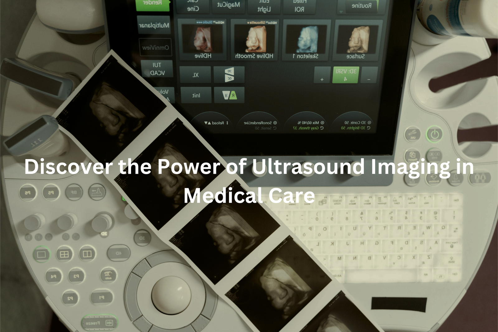
Ultrasound Imaging uses sound waves to give doctors a peek inside your body. Find out how it works, what to expect, and why it’s safe.
Ultrasound imaging is a way for doctors to see inside your body using sound waves instead of X-rays. This method helps them check soft tissues, blood vessels, and even look at babies developing in the womb. During a scan, a special device sends out sound waves and captures the echoes to create images.
It’s painless and safe since it doesn’t use harmful radiation. You’ll probably hear some funny sounds while the machine works. If you’re curious about how ultrasound helps in medicine, keep reading to find out more!
Key Takeaway
- Ultrasound imaging uses high-frequency sound waves to create pictures of the inside of the body.
- It’s a safe method widely used for pregnancy ultrasounds and other medical checks.
- Preparing for an ultrasound is easy and usually involves just a few simple steps.
Ultrasound Scans
The first time seeing an ultrasound machine in action was a bit of a revelation. What looked like a simple remote control turned out to be capable of seeing right through skin and muscle—using just sound waves. It’s a pretty clever piece of tech, really.
Here’s how it works: the machine sends out high-pitched sound waves, about 2 to 18 megahertz (that’s way beyond a dog’s hearing range). These waves bounce off the insides, kind of like sonar, and create an image on the screen.
While most folks think ultrasound is just for checking on babies, it’s actually used for so much more. Doctors use it to:
- Watch hearts beat
- Track blood flow
- Check on kidneys, livers, and other organs
- Detect lumps or cysts
The best part? There’s no radiation or needles involved—just some cold gel and a probe that slides across the skin. Sometimes, you’ll need a full bladder for better imaging (not the most fun experience), but it beats surgery any day. A quick pro tip: wear comfy clothes. Those exam tables aren’t exactly five-star comfort.
Pregnancy Ultrasound
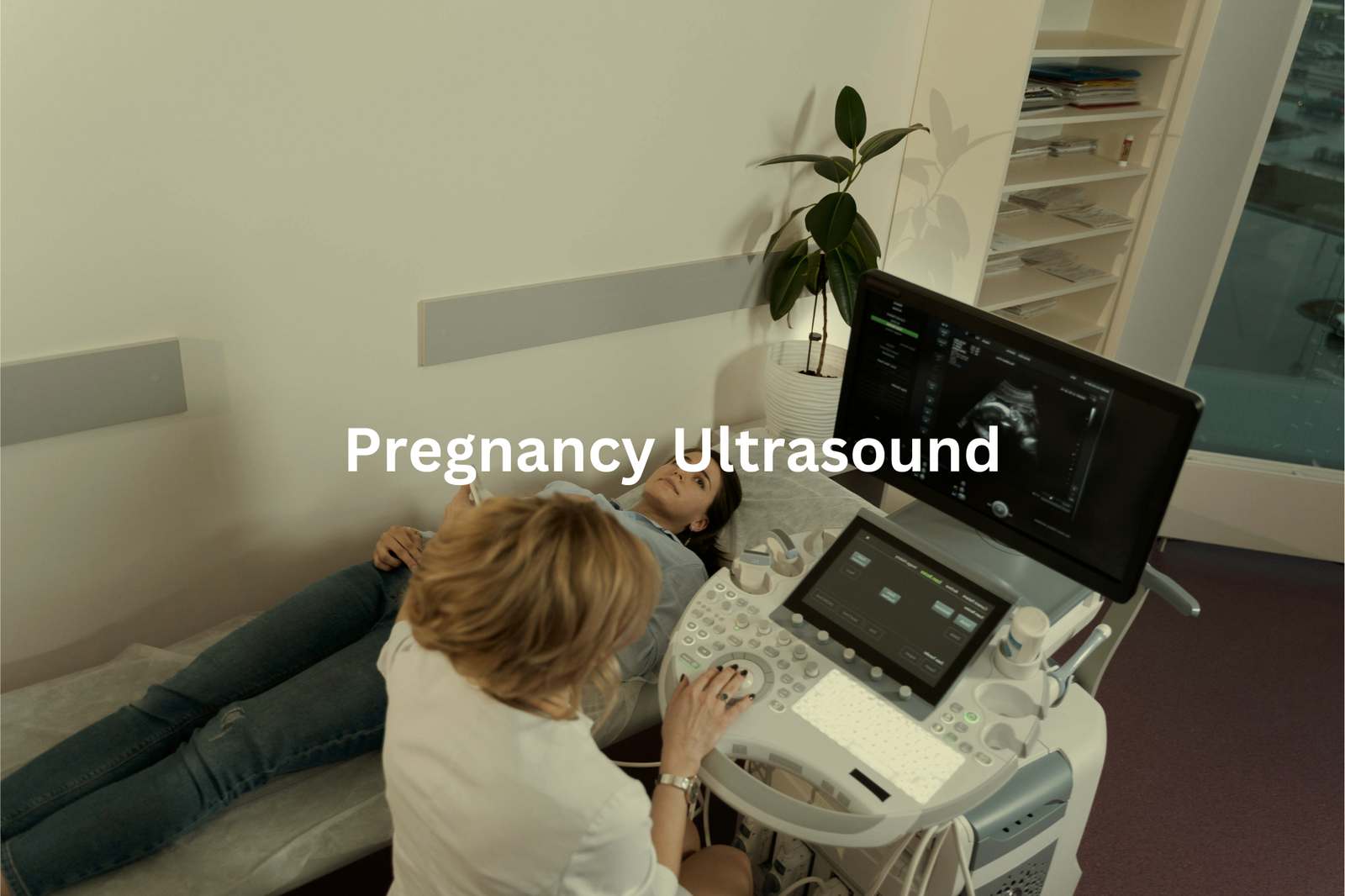
Ultrasound scans are pretty incredible, using sound waves between 2-18 MHz to create images of the inside of the body. (1)
Pregnancy scans, in particular, are a big part of the process (though they’re not just for babies). The early scans help doctors figure out the baby’s growth and health, starting with:
- First scan (6-12 weeks): Checks if everything’s going as planned
- NT scan (11-14 weeks): Looks at fluid behind the baby’s neck
- Big scan (18-22 weeks): Examines all the important parts
- Growth checks: Monitors baby’s development
One tip I’ve picked up? Drink about 500ml of water an hour before the scan. It helps improve visibility. And don’t stress if the sonographer points at a grey blob you can’t quite identify—those images can be tricky to read at first!
It takes a bit of practice, but once you’ve seen a few, they start to make sense. Keep it comfy—those exam tables aren’t exactly built for lounging.
Doppler Ultrasound
Doppler ultrasound is a clever tool. Unlike regular ultrasound, which just takes pictures, Doppler listens to blood as it moves. The machine sends sound waves that bounce off the blood cells, and when they return, they help form a picture doctors can read. Faster blood flow makes higher-pitched sounds, and slower flow creates lower ones. (2)
Doctors often use Doppler ultrasound for a few key things:
- Monitoring blood flow to a baby during pregnancy
- Checking for heart issues
- Finding blood clots in the legs or arms
The process itself is fairly simple. A probe covered in cold gel slides across the skin. It might feel a little uncomfortable at times, especially if the sonographer needs to press harder to get deeper images. But it’s generally painless.
One thing to keep in mind is that some scans need a full bladder for better visibility. It’s not the most fun part of the procedure, but it’s much less invasive than the alternatives. For anyone getting this scan, the best advice is to stay relaxed, wear something comfortable, and know that you won’t need any recovery time once it’s done.
How Ultrasound Works
Ultrasound machines are oddly simple-looking. A computer and a small probe, and somehow they can peer inside the body. It’s like sound does the heavy lifting. The machine sends waves out, way beyond what we can hear, and listens for echoes bouncing back. These echoes turn into a black-and-white image, where fluids show up dark and bones look bright.
Doctors use ultrasound for quite a bit. It helps with:
- Monitoring a baby’s development
- Checking the heart’s rhythm
- Spotting problems in organs like kidneys
The whole procedure usually takes around 30 minutes. A cold gel gets spread across the skin to help the probe slide easily. There’s no radiation, no needles. It’s a simple process, yet it provides so much information.
A tip? If you’re having an ultrasound, make sure to wear comfy clothes. Sometimes you’ll need a full bladder for a clearer picture (not the most pleasant thing, but worth it).
Ultrasound Preparation
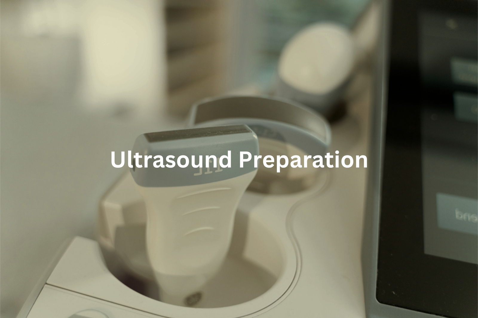
Ultrasound prep is a game of patience, especially when it comes to a full bladder. It’s not hard to get ready for a scan, but timing’s key. For a pelvic ultrasound (like checking the bladder or early pregnancy), drink about 750mL of water an hour before. That’s roughly three cups. You need the bladder full enough for the sound waves to travel clearly.
For a tummy scan (liver, kidneys, gallbladder), things get a bit trickier. No food or drink except water for 6-8 hours before. Learned that the hard way when a simple breakfast messed up my first scan. The bubbles from digestion made the whole thing blurry.
One tip? Don’t overdo the water. You want a full bladder, but not bursting. And if you’re unsure about the instructions, give the clinic a ring. Better safe than squirming.
Ultrasound Safety
Ultrasound might sound scary at first, but it’s actually a simple and safe process. It uses sound waves, not X-rays (which is a big plus for safety). The machine sends high-frequency sound waves through the body, and they bounce back to create real-time pictures. No radiation involved, just a fancy version of a bat’s sonar. (3)
Some key points to remember:
• It’s completely painless
• Shows moving pictures in real-time
• Uses sound, not X-rays
• Safe enough for babies
A warm gel gets spread on the skin, and a small device called a transducer is used to collect the images. It usually takes about 20 minutes, and the pictures pop up right there on the screen. It’s a pretty cool process, really.
While ultrasound isn’t something you’d do for fun, there’s nothing to worry about when you actually need one. It’s straightforward and comfortable enough for almost anyone.
Ultrasound Uses
Every time I walk past the ultrasound room at Yale-New Haven Hospital during my clinical Ultrasound isn’t just for baby scans. It’s a quiet, unassuming powerhouse in medicine. The machine uses sound waves—well above what we can hear (around 20,000 Hz)—to capture images inside the body. The probe moves like an artist’s brush, painting pictures on the screen in black and grey.
Ultrasound can check on all sorts of things, like:
- A beating heart
- Blood flowing through veins
- Organs, from kidneys to thyroid
- Lumps or swelling that don’t feel quite right
The best part? No radiation, no dyes, just instant results. It’s like shining a flashlight inside without the light. The pictures show up while the probe’s still moving, and no waiting is needed for the results. When the body is involved, timing matters, and ultrasound gets it right.
Vascular Ultrasound
I watched my friend’s dad get a vascular ultrasound last month, and it really showed me how amazing these scans are. His leg was all puffy and red, and he couldn’t walk right. The The ultrasound found a blood clot hiding behind the knee—dangerous stuff if it hadn’t been spotted.
The test’s pretty straightforward. The doctor uses a probe with cold gel, then watches as blood flows through the veins on a screen. Red and blue colours light up, showing the direction of the flow. If there’s a blockage, it’s easy to see.
Ultrasounds can check for:
- Blood clots in veins
- Blocked arteries
- Abnormal blood flow patterns
No needles, no radiation, just gel and a quick 15-minute scan. After the scan, the right treatment—like blood thinners—can keep things on track. For anyone with sore, swollen legs, an ultrasound could save a lot of trouble.
Abdominal Ultrasound
Ultrasound’s a strange thing. The way sound waves turn into pictures of what’s happening inside without a single incision. It’s pretty mind-blowing.
Doctors often reach for an ultrasound when someone’s belly’s giving trouble. It doesn’t hurt, no needles or radiation—just some gel and a handheld scanner (called a transducer, if you’re curious).
What can it check for?
- Liver problems – bright, patchy if there’s fat buildup
- Gallstones – show up as tiny white marbles
- Kidney issues – stones or blockages
- Pancreas – tricky but possible to see
Here’s a tip: skip breakfast before your scan. An empty stomach (8-12 hours) gives the best results. Nothing worse than having to reschedule because someone grabbed their morning coffee. If sharp pain hits after a greasy meal, it might be gallbladder trouble—time for a scan.
Ultrasound Results
Ultrasound results don’t take long to show up. Usually, about 24 to 48 hours. The process is simple, really. First, a radiologist reviews the images. Then, the doctor gets the report. After that, they’ll call or book an appointment to discuss the findings.
The machine itself uses sound waves—about 20,000 Hz—way beyond what we can hear. It’s pretty amazing how it turns sound into a picture of what’s going on inside.
Some common things doctors look for:
- Liver issues – signs of fatty buildup
- Gallstones – little white spots
- Kidney problems – blockages or stones
One thing that might help: write down questions before your follow-up appointment. It’s easy to forget once you’re sitting there, waiting to hear what’s next. If something hurts in the abdomen after a fatty meal, it might be time to get checked.
FAQ
What is ultrasound imaging?
Ultrasound imaging, also known as medical ultrasound, is a diagnostic technique that uses high-frequency sound waves to create images of structures inside the body. These sound waves bounce off the tissues and organs, and the reflections are captured by a special device to produce real-time images.
How does ultrasound work?
Ultrasound imaging works by sending high-frequency sound waves into the body using a special probe. These sound waves bounce off the different tissues and organs, and the reflections are captured by the probe to create an image. The speed of sound through the body is used to determine the location and size of the structures being imaged.
What can ultrasound be used for?
Ultrasound imaging can be used to examine a wide range of body parts, including the heart, blood vessels, kidneys, liver, and reproductive organs. It is particularly useful for monitoring pregnancy, as it can provide detailed images of the developing fetus. Ultrasound is also used to guide certain medical procedures, such as biopsies and injections.
What are the benefits of ultrasound?
One of the main benefits of ultrasound is that it is a non-invasive and painless procedure. It does not use ionising radiation, like X-rays or CT scans, which makes it a safer option for many patients. Ultrasound is also relatively affordable and can provide real-time images of the body, which can be useful for diagnosing and monitoring various medical conditions.
How do I book an ultrasound appointment?
To book an ultrasound appointment, you can contact your local medical imaging provider or healthcare provider. Many providers offer online booking options, as well as appointments on Fridays and Saturdays to accommodate busy schedules. Be sure to check if bulk billing is available, which can help reduce the cost of the scan.
What should I expect during an ultrasound?
During an ultrasound, you will be asked to lie down on an examination table, and a clear gel will be applied to the area being examined. The technician will then move a small, handheld probe called a transducer over the area, which sends high-frequency sound waves into the body. The reflections of these sound waves are used to create real-time images that can be viewed on a monitor.
What is the difference between transvaginal and transabdominal ultrasound?
Transvaginal ultrasound involves inserting a probe into the vagina to obtain images of the reproductive organs, such as the uterus and ovaries. This type of ultrasound can provide more detailed images than a transabdominal ultrasound, which involves placing the probe on the abdomen. Transvaginal ultrasound is commonly used during pregnancy to assess the developing fetus.
How do I prepare for an ultrasound?
Preparation for an ultrasound can vary depending on the type of scan being performed. For some scans, you may need to drink a certain amount of water beforehand to ensure a full bladder. For others, you may need to fast for a period of time. Your healthcare provider will provide specific instructions on how to prepare for your ultrasound appointment.
What is the difference between ultrasound and other imaging techniques?
Ultrasound is different from other imaging techniques, such as X-rays and CT scans, in that it uses high-frequency sound waves instead of ionising radiation. This makes ultrasound a safer option, particularly for pregnant women and children. Ultrasound can also provide real-time images and is generally more affordable than other medical imaging services.
Conclusion
Ultrasound imaging is a safe and useful way to look inside your body. It helps doctors see how babies are growing, check blood flow, and find different health issues. If you need an ultrasound, it’s important to follow the preparation steps, which might include drinking water before the test. Knowing what to expect can make the experience easier. So, when it’s time for your appointment, you’ll feel ready and confident with the process ahead.
References
- https://www.sufw.com.au/services/pregnancy-ultrasound/
- https://www.healthywa.wa.gov.au/Articles/A_E/Doppler-ultrasound
- https://www.healthdirect.gov.au/ultrasound