
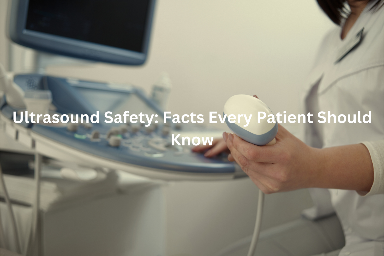
Curious about ultrasound safety? Find out how these sound waves work safely to show what’s inside your body, especially for expectant mums.
Ultrasound safety is really important. Ultrasounds use sound waves, which are safe and do not hurt you or your baby. Doctors say that when done properly, ultrasounds are very safe. They help see inside the body without using x-rays or other harmful stuff. Pregnant women often have ultrasounds to check on their unborn babies.
It’s good to ask your doctor any questions you have about the procedure. Knowing how it works can help you feel better. So, if you’re curious about ultrasounds, keep reading to learn more about their safety and why they’re used.
Key Takeaway
- Ultrasounds do not use harmful ionising radiation, making them safer than some other imaging tests.
- Following safety guidelines helps minimise any risks associated with ultrasound exams.
- Always talk to your healthcare provider if you have questions or concerns about ultrasound safety.
What is an Ultrasound?
Ultrasound scans, commonly known as sonograms in Australia, create detailed images of unborn babies through sound waves that bounce off the developing fetus. The technology works much like sonar used to map ocean floors.
Australian pregnancy care includes two main scans. The first dating scan happens between 8-12 weeks, pinpointing how far along the pregnancy is. The second major scan occurs at 18-22 weeks, checking the baby’s growth and development (measuring everything from head size to leg length).
During these scans, sonographers use a transducer – a special wand-like device – covered in conductive gel. The device sends sound waves through the mother’s abdomen, creating black and white images on a nearby screen. Sometimes babies turn away or cover their faces with tiny hands, making the scan take longer.
These medical scans serve as vital health checks, spotting potential concerns early while giving parents their first glimpse of their growing baby.
Why is Ultrasound Considered Safe?
Ultrasound scans create pictures using sound waves that bounce off different body parts, much like echoes in a cave. The process starts with cold gel on the skin (about 20 degrees Celsius), which helps the sound waves travel better.
Australian healthcare makes ultrasound accessible through Medicare benefits. The procedure costs between $60-200, with rebates covering up to 85% of the fee at approved clinics.
These scans work differently from X-rays:
- No radiation exposure
- Real-time moving images
- Pain-free process
- Quick results (15-30 minutes)
The Royal Australian and New Zealand College of Obstetricians and Gynaecologists confirms ultrasounds are safe for both mother and baby. The high-frequency waves (between 2-15 MHz) can’t be heard by human ears but create detailed images of internal structures(1).
Patients should wear comfortable clothes that allow easy access to the scan area. The gel might feel cold at first but warms up quickly on contact with skin.
What are the Guidelines for Ultrasound Safety?
Sources: Clinical Imaging.
The whoosh-whoosh sound of a baby’s heartbeat fills ultrasound rooms across Australia daily. These sound waves, operating at frequencies between 2-15 MHz, create images that guide medical professionals through pregnancy journeys.
Australian healthcare maintains strict regulations for ultrasound use. The Australasian Society for Ultrasound in Medicine (ASUM) oversees these practices, ensuring only qualified sonographers and doctors operate the equipment. They follow the ALARA principle (As Low As Reasonably Achievable), which keeps power settings minimal while maintaining image quality.
Unlike X-rays that use radiation, ultrasound technology works through harmless sound waves. Medical guidelines in Australia recommend:
• Scans only when medically necessary
• Qualified professionals performing all examinations
• Standard 2D scans over elective 4D imaging
Medical centers across the country, from Sydney to Perth, stick to these safety protocols. The focus remains on getting clear diagnostic images while keeping both mother and baby safe throughout the process.
Are There Any Risks?
Ultrasound machines, those boxy devices found in medical centers across Australia, work like underwater sonar to create images of the body’s internal structures. The equipment sends out high-frequency sound waves (between 2-15 MHz) that bounce off organs and tissues.
Medical professionals in Australian hospitals follow strict guidelines from ASUM (Australasian Society for Ultrasound in Medicine) when performing these scans. The process needs careful attention to three key areas:
• Power output settings must stay within safe limits
• Scan duration should be as brief as needed
• The probe requires proper positioning
While ultrasound scanning is generally safe, the sound waves can cause tiny temperature increases (usually less than 1°C) in body tissues. Medical staff apply the ALARA principle – using the lowest possible power level to get clear images.
Unlike X-rays or CT scans, ultrasound doesn’t use radiation. Still, patients should only get scans when medically necessary, and always from certified sonographers or radiologists.
How is Infection Controlled?
The distinct scent of medical-grade disinfectant and hand sanitiser fills every ultrasound room in Australian hospitals. This smell signals a clean, safe environment for medical imaging procedures.
At leading medical centres across Australia, sonographers follow strict cleaning protocols before each ultrasound scan:
• Opening new, sealed ultrasound gel packets
• Disinfecting the ultrasound probe with hospital-grade wipes
• Using fresh gloves for each patient
• Recording cleaning times and dates
The ultrasound probe requires thorough sanitisation between patients (this small device makes contact with the skin during scans). The Australasian Society for Ultrasound in Medicine (ASUM) emphasises these cleaning standards, particularly for internal examinations where infection risks increase(2).
Patients can watch these safety steps during their visits. Medical staff should demonstrate the cleaning process when asked. Speaking up about hygiene concerns helps maintain high safety standards in medical settings.
What Should Patients Consider?

Ultrasound scans at medical centers across Australia help doctors see inside the body without any cuts or pain. The process takes about 20 minutes, and patients simply lie on an examination bed while a trained sonographer works the scanning equipment.
The procedure starts with clear, water-based gel applied to the skin. This gel might feel cool at first, but it helps the scanning wand move smoothly across the area being checked. The machine uses special sound waves (between 1-15 MHz) to create pictures of organs and tissues.
Before the scan, patients should:
- Wear loose, comfortable clothing
- Tell staff about any allergies
- Follow specific preparation instructions
- Ask questions if unsure about anything
Most people describe the sensation as gentle pressure on their skin. The sonographer moves the scanning wand carefully to get the best possible images for the doctor to review. Medicare covers many ultrasound services at approved clinics throughout Australia.
Who Can Perform an Ultrasound?
Ultrasound scans at Royal Prince Alfred Hospital show patients what’s happening inside their bodies. The machine looks like a regular computer with a special wand attachment, but it uses smart technology to create images.
During an ultrasound:
- The sonographer applies cool gel to the skin
- A handheld device (15 cm in length) moves across the area
- Sound waves between 2-15 MHz bounce off internal structures
- Black and white images appear on the screen
Australian sonographers, the medical professionals who perform ultrasounds, guide patients through the process. The procedure takes about 20 minutes and causes no pain. The gel might feel a bit cold at first, but that’s normal.
The technology works like an underwater sonar system, sending sound waves through body tissue to create detailed pictures. Patients who feel anxious can speak up – sonographers perform these scans hundreds of times each week and know how to make the experience comfortable.
What Happens After an Ultrasound?
Patients at Sydney’s Royal North Shore Hospital often wait for their ultrasound scans in a quiet room. The process follows a simple path that helps keep everyone informed about their health.
During an ultrasound scan (which uses sound waves at 1-15 MHz), medical staff capture detailed images of the body. The procedure typically takes 20-30 minutes, and patients stay still while the sonographer works.
After the scan, these steps follow:
- The radiologist reviews all images thoroughly
- Results get shared with patients if everything looks normal
- Further tests might be needed if the doctor spots anything unusual
Medicare covers most ultrasound costs at Australian hospitals, though some places might have extra fees. Patients should:
- Bring their Medicare card
- Wear loose-fitting clothes
- Ask questions if something isn’t clear
- Follow any preparation instructions given before the scan
The medical team at Royal North Shore explains results in clear, simple terms, making sure patients understand their situation.
Where to Get an Ultrasound?
Getting an ultrasound scan in Australia is a straightforward medical procedure. Medicare covers most diagnostic imaging services across the country, making them accessible to many people.
Several types of facilities offer ultrasound services:
- Public hospitals (like Royal Prince Alfred)
- Private medical centres
- Specialist imaging clinics
The process starts with a GP referral. Patients need to:
- Book an appointment
- Bring their Medicare card
- Follow any preparation guidelines
- Wear comfortable clothing
Most suburban areas have imaging centres within 10 kilometres (about 6.2 miles), making them easily reachable. The radiologists at these facilities use high-frequency sound waves (between 2-15 MHz) to create detailed images of organs and structures inside the body.
Booking an ultrasound typically takes 24-48 hours, though some centres offer same-day appointments. The scan itself usually lasts 15-30 minutes, depending on the area being examined.
Why is Understanding Ultrasound Safety Important?
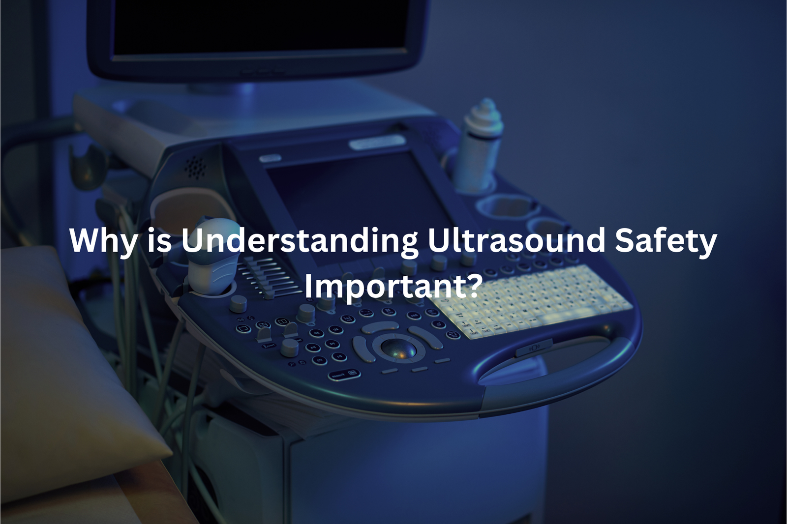
Ultrasound scans help doctors see inside the body without using harmful radiation. These scans use sound waves (above 20,000 Hz) to create clear pictures of organs, tissues, and growing babies.
During an ultrasound scan, patients lie on a bed while a sonographer applies a cool gel to their skin. The gel helps the transducer (a small handheld device) slide smoothly across the area being examined. The transducer sends sound waves into the body and catches their echoes to make detailed images on a screen.
Common uses for ultrasound include(3):
• Checking baby growth during pregnancy
• Looking at heart function
• Examining muscles and joints
• Finding kidney stones
• Checking blood flow
A typical ultrasound takes about 30 minutes. The procedure doesn’t hurt, and patients can ask questions throughout the scan. Most medical centres encourage questions, as understanding the process helps reduce anxiety about the examination. Sonographers recommend wearing loose, comfortable clothing to these appointments.
FAQ
What are the MI and TI values in an ultrasound exam, and why are they important?
The MI (Mechanical Index) and TI (Thermal Index) values provide important information about the sound energy used during an ultrasound exam. The MI measures the risk of physical damage to tissues, while the TI estimates the potential for temperature increase in the body. Clinicians use these values to ensure the ultrasound beam is operating at safe levels and not causing any harmful effects to the patient’s soft tissues, blood vessels, or internal organs.
How can Doppler ultrasound be used to evaluate blood flow in the body?
Doppler ultrasound technology uses high-frequency sound waves to detect and measure the movement of blood through the body’s blood vessels. This allows clinicians to assess blood flow, identify any blockages or abnormalities, and monitor conditions like peripheral artery disease, deep vein thrombosis, and fetal heart rate. Doppler ultrasound is a valuable tool in both diagnosis and ongoing patient care.
What is the purpose of 3D and 4D ultrasound imaging?
3D and 4D ultrasound scans provide a more detailed, three-dimensional view of the internal structures being examined, such as the fetus, internal organs, or breast tissue. 4D ultrasound adds the dimension of time, allowing the clinician to see movement and function in real-time. These advanced imaging techniques can help identify potential issues early and guide medical decision-making, particularly in obstetric and diagnostic applications.
How can ultrasound be used to detect potential issues in early pregnancy?
Ultrasound is an essential tool for monitoring fetal development and identifying any potential problems in early pregnancy. It can be used to determine gestational age, check the health of the embryo or fetus, assess amniotic fluid levels, and evaluate the condition of the umbilical cord and placenta. Clinicians can also use ultrasound to screen for chromosomal abnormalities and other congenital issues, allowing for early intervention and management.
What precautions are taken to ensure patient safety during an ultrasound exam?
Clinicians follow strict safety protocols when performing ultrasound exams to protect patient health. This includes carefully monitoring the acoustic output, avoiding unnecessary exposure, and obtaining informed consent. The use of contrast agents or Doppler techniques is also closely regulated to minimise any potential risks. Patients are encouraged to discuss any concerns with their healthcare provider to ensure a safe and comfortable ultrasound experience.
Conclusion
Ultrasound safety really matters when we use this important tool for checking health. Ultrasounds are mostly safe because they don’t use harmful radiation like X-rays do. It’s a bright idea to stick to safety guidelines and talk with your doctors. They can help make sure everything goes smooth and safe. If you’re curious about how ultrasounds work or if they’re safe for you, just ask your doctor. They’re there to help answer your questions!
References
- https://ultrasoundcare.com.au/debunking-ultrasound-myths/
- https://www.nanosonics.com.au/infection-prevention/infection-risk-from-ultrasound/
- https://www.healthywa.wa.gov.au/Articles/U_Z/Ultrasound


