
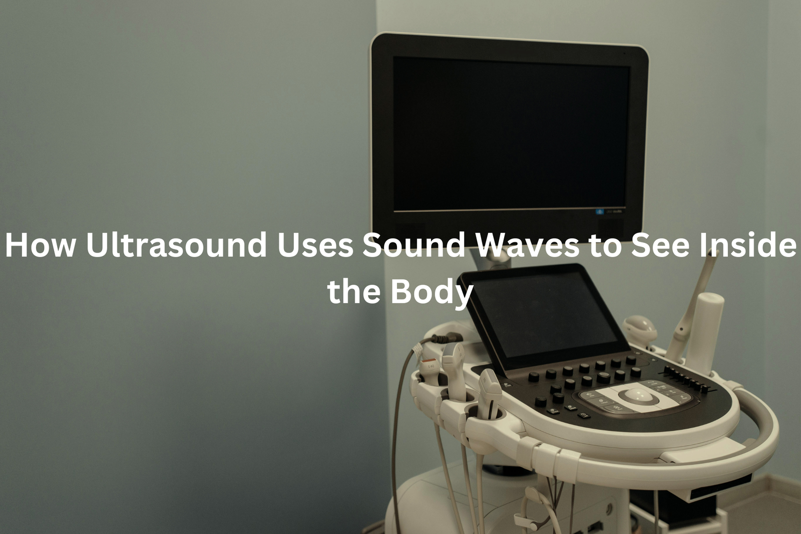
Ultrasound uses sound waves to create pictures inside our bodies. Discover how this non-invasive tool helps doctors see what’s happening without surgery!
Ultrasound technology creates detailed pictures of the body’s inner workings through sound waves (frequencies above 20,000 Hz). The non-invasive scan bounces these waves off organs and tissues, building clear images on a screen. Medical professionals use ultrasounds daily for various checks. During pregnancy scans, they spot baby movements and measure growth.
The technology also finds gallstones, examines hearts, and checks liver function. The procedure takes 15-45 minutes, depending on the area being scanned. Patients lie still while a sonographer moves a small probe across their skin with gel. No radiation means it’s safe for repeated scans, making it a standard choice in Australian healthcare centres.
Key Takeaway
- Ultrasound can check many body parts, like the heart and belly, using sound waves.
- It helps doctors see babies growing in their mummies and find out if they are healthy.
- Ultrasound is safe and does not use harmful rays like some other tests.
What is Ultrasound?
Ultrasound scans let doctors see inside the body using sound waves that humans can’t hear (above 20,000 cycles per second). The process works a bit like sonar on submarines, sending waves through tissue and creating pictures from the echoes.
During an ultrasound scan, patients lie on a bed while a sonographer spreads cool gel on their skin. The gel helps sound waves travel better between the transducer (the scanning tool) and the body. The whole thing takes about 30 minutes, and patients don’t feel any pain.
The machine shows everything in real-time on a screen, kind of like a special medical TV. Unlike X-rays, ultrasounds don’t use any radiation, making them safe for regular check-ups and pregnancy scans.
Common ultrasound uses:
- Checking baby development
- Looking at muscles and joints
- Examining organs like the heart or liver
- Finding blood flow problems
Patients who want to understand more can ask their sonographer to explain the grey and white images on screen.
Uses for Babies
Ultrasound scans use sound waves (similar to those used by dolphins) to create images of growing babies inside the womb. These scans work like an underwater camera, showing detailed pictures of the developing baby.
From 6 weeks, doctors can detect a tiny heartbeat flickering on screen. By 12 weeks, babies measure about 6 centimetres and perform gentle movements in the amniotic fluid. The scan reveals babies swimming, sleeping, or moving around in their watery home.
During ultrasound checks, medical professionals look for:
- Baby’s measurements from head to bottom
- Heart rate and activity
- Baby’s position
- Number of babies present
At the 20-week scan, babies might show different activities like yawning or stretching. While mothers mightn’t feel these movements yet, the ultrasound screen displays them clearly. For best scan results, patients shouldn’t drink excessive water beforehand – modern ultrasound machines don’t need a full bladder for clear images.
Looking at the Heart
An echocardiogram works like a window into the human heart(1). The ultrasound machine creates moving pictures using sound waves (at frequencies between 2-10 MHz), showing every pump and valve in real-time.
During the scan, a medical professional applies gel to the patient’s chest and moves a small device called a transducer across the area. The procedure doesn’t cause any pain, though patients might notice the cool sensation from the gel.
This non-invasive test helps doctors check:
- Heart chamber size and shape
- Valve function and movement
- Blood flow patterns
- Muscle wall strength
- Overall heart performance
The scan takes about 30-45 minutes, and patients can breathe normally throughout. Unlike x-rays, echocardiograms don’t use radiation, making them safe for repeated tests. Most Australian medical centres and hospitals offer this diagnostic tool, which Medicare typically covers with a proper referral from a GP. Patients should wear comfortable, loose clothing and might need to remove items from their chest area during the procedure.
Checking Muscles and Joints
Ultrasound imaging helps doctors see inside sports injuries without surgery(2). The machine uses sound waves (between 1-20 MHz) to create moving pictures on a screen, similar to a black-and-white movie of the body’s inner workings.
While many know ultrasound from pregnancy scans, it’s also a valuable tool for checking sports injuries. The technology shows:
• Swelling in soft tissues
• Inflamed tendons
• Muscle tears and bruising
• Joint problems
During an exam, patients lie still as the doctor moves a small device called a transducer over the injured area. The transducer sends sound waves through the skin, creating clear images of what’s happening underneath. Most scans take about 15-20 minutes.
Athletes with ongoing pain should get checked early. Quick treatment often means faster healing, and ultrasound helps doctors spot problems that might not show up in a regular exams better. Ultrasound can show what’s wrong straight away, which helps doctors fix it properly.
Understanding Blood Flow
Sources: RVT Pro.
Doppler ultrasound scans help doctors spot blood flow problems in patients’ legs without any cuts or needles. This clever medical tool (using sound waves at frequencies between 2-18 MHz) shows blood movement through veins and arteries right on a screen.
During the scan, medical staff apply a cool gel to the skin and move a small wand across the area. The whole process takes about 30 minutes, and patients can watch their blood flow patterns appear in real-time.
The test checks for several issues:
• Blocked blood vessels
• Heart pumping efficiency
• Poor circulation
• Blood clots
Many Australians with diabetes or high blood pressure get these scans regularly. Medicare covers most of the cost at public hospitals, though private clinics might charge extra fees. Anyone noticing swelling or pain in their legs should ask their GP about getting a Doppler ultrasound. Early detection makes treating circulation problems much easier.
Helping with Procedures
Ultrasound-guided biopsies at Royal North Shore Hospital in Sydney take about 30 minutes from start to finish, with the actual procedure lasting just 15 minutes. A doctor uses specialised equipment – a transducer connected to a computer screen – to display black and white images of the body’s internal structures.
The procedure offers several benefits:
- Real-time imaging of organs and tissues
- Clear views of blood vessels to avoid complications
- Precise needle placement for accurate sampling
During the procedure, patients receive local anaesthetic to minimise discomfort. The medical team at Royal North Shore Hospital performs roughly 20 ultrasound-guided biopsies weekly (making it a routine procedure in their clinic).
The technology makes biopsies safer and more accurate than traditional methods. Medical professionals can watch the needle’s path on screen throughout the entire process, ensuring they collect tissue samples from the correct location while avoiding sensitive areas.
For Kids

Ultrasound scans at children’s hospitals bring smiles instead of tears. The process starts with warm gel (about 35 degrees Celsius) spread across the skin, followed by a small handheld device that looks like a remote control.
These scans, performed by qualified sonographers, use sound waves to create pictures of what’s inside the body. The kidneys, measuring roughly 10 centimetres in length, appear as bean-shaped structures on the black-and-white screen.
Children’s ultrasound appointments typically include(3):
- A 15-20 minute scanning time
- Zero radiation exposure
- No needles or painful procedures
- Clear images for medical teams
- A printed image to take home
The Royal Children’s Hospital Melbourne, one of Australia’s leading paediatric centres, makes these scans child-friendly. The sonographers show children the moving images on screen, making the experience more like watching a special kind of TV show than having a medical test. For most kids, getting an ultrasound feels as simple as having their photo taken.
Sonographers: The Ultrasound Helpers
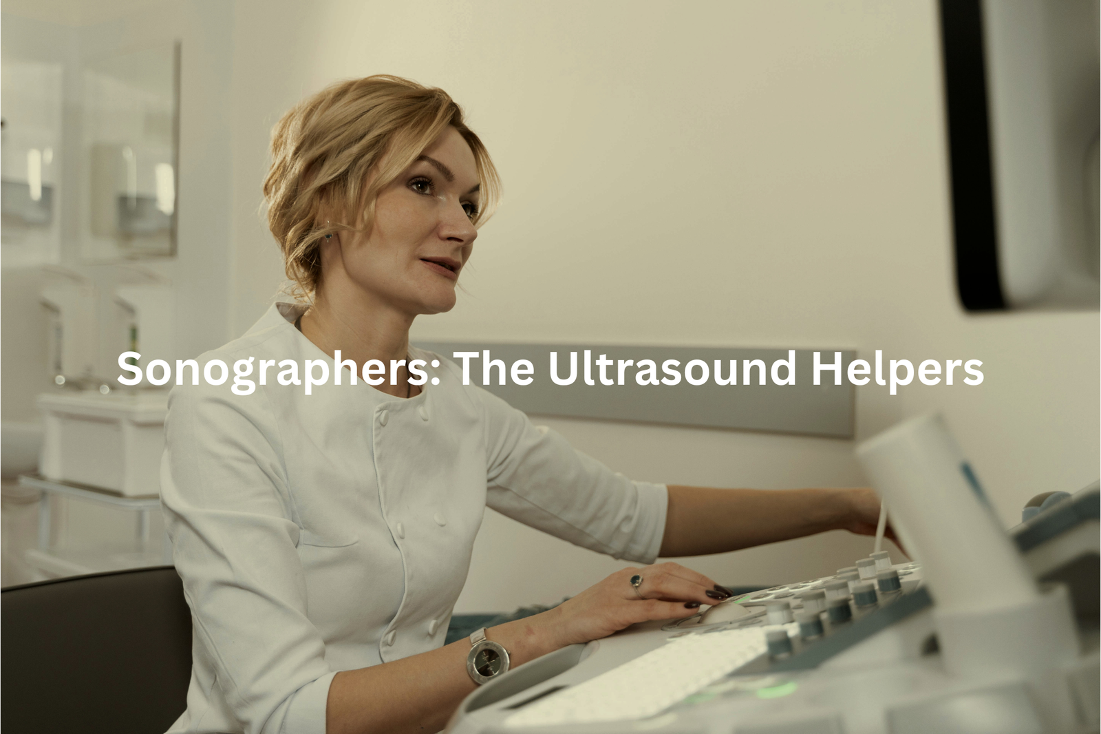
Sonographers work like skilled photographers of the human body, using special wands called transducers to capture images inside patients. At hospitals across Australia, these medical professionals spend their days looking at black-and-white pictures that show everything from tiny hearts to growing babies.
Getting into this field takes dedication – a 4-year university degree plus extra certifications (through the Australian Sonographer Accreditation Registry). The job needs steady hands and sharp eyes to spot potential health issues.
Their daily tasks include:
• Prenatal checkups
• Musculoskeletal scans
• Cardiac imaging
• Organ function tests
Each scan produces detailed images that help doctors make better decisions about patient care. Modern ultrasound machines can show blood flow patterns and create 3D images of organs, making them essential tools in Australian healthcare. While they might not wear the traditional white coats, sonographers play a crucial part in finding health problems early.
No Harmful Rays
Ultrasound scans, a common sight in Australian hospitals, use sound waves at 20,000 vibrations per second to create detailed images of the body’s inner workings. Unlike X-rays or CT scans, these sound waves don’t use any radiation, making them completely safe for patients of all ages.
The process is straightforward in medical settings. A sonographer applies a special gel to the skin and moves a handheld probe across the area. The entire scan typically takes 15-20 minutes, and patients can watch the images appear on screen in real-time.
Key benefits of ultrasound technology:
• Non-invasive and painless
• Shows movement of organs like the heart
• Safe for repeated examinations
• Provides instant results
• Cost-effective compared to other imaging methods
Since its introduction to Australian healthcare in the 1950s, ultrasound has become a trusted diagnostic tool. The technology continues to advance, offering clearer images while maintaining its reputation as one of the safest medical tests available.
FAQ
What is a CT scan and how does it differ from ultrasound?
A CT scan uses X-rays to create detailed images of the body’s internal structures, while ultrasound uses high-frequency sound waves to capture real-time images. CT scans are better for viewing bone and detecting conditions like cancer, while ultrasound is preferred for examining soft tissues, fluids, and the heart.
How can ultrasound be used in physical medicine and rehabilitation?
Ultrasound has a wide range of uses in phys med, such as imaging muscles, tendons, and joints to diagnose injuries. It can also guide injections and measure blood flow and heart rate. Ultrasound helps physical therapists monitor patient progress and tailor treatments for conditions like muscle tears, joint problems, and vascular disorders.
What is “real-time” ultrasound and how is it used?
Real-time ultrasound allows doctors to see internal body parts in motion, like the heart beating or blood flowing. This helps them assess organ function, fluid levels, and other dynamic processes. Real-time imaging is particularly useful for guiding procedures like biopsies and injections, as the doctor can see the instrument’s movement.
Where can I find more information on ultrasound technology?
For in-depth technical details on ultrasound, try resources like CRC Press books, IEEE Transactions on Biomedical Engineering, and the Acoustical Society of Australia journal. These cover the physics, engineering, and medical applications of acoustic waves, imaging modes, and related technologies like Doppler and 3D/4D imaging.
How does the speed of sound in soft tissues affect ultrasound imaging?
The speed of sound varies slightly in different soft tissues, which can cause distortions in the image. Factors like the body part, fluid content, and patient’s age influence the speed. Compensating for these variations is important for achieving accurate, high-quality ultrasound images of organs, muscles, blood vessels and other structures.
What are the benefits of using linear arrays vs. phased arrays in ultrasound?
Linear array transducers provide a wide field of view and high-resolution images, making them useful for examining surface structures like the muscles and tendons. Phased arrays, on the other hand, can steer the sound beam to image deeper structures like the heart and abdomen. The choice depends on the clinical application and the part of the body being imaged.
How does Doppler ultrasound work and what is it used for?
Doppler ultrasound uses the Doppler effect to detect movement, like blood flowing through vessels or a fetus’s heartbeat. This allows doctors to measure the speed and direction of fluid flow, which is valuable for assessing vascular health, organ function, and fetal development. Doppler is a key tool for diagnosing conditions affecting the heart, blood vessels, and urinary tract.
Conclusion
Ultrasound technology serves as a vital medical tool in Australian healthcare. The device sends high-frequency sound waves through the body, which bounce back to create detailed images of organs and tissues (operating at frequencies above 20,000 Hz). Medical professionals use these scans to examine unborn babies, check heart function, and spot potential health issues. The non-invasive nature of ultrasounds makes them a safe choice for diagnostic imaging in clinics and hospitals across the country.
References
- https://www.svhhearthealth.com.au/procedures/imaging/echocardiogram-echo
- https://www.gehealthcare.com.au/products/ultrasound/point-of-care-ultrasound/musculoskeletal-ultrasound
- https://www.childrens.health.qld.gov.au/health-a-to-z/ultrasound


