

X-ray uses in medicine are vital! Learn how these scans help doctors see inside our bodies, diagnose issues, and plan treatments quickly and safely.
X-rays are super important in medicine. They let doctors peek inside our bodies without needing surgery. These machines take pictures of bones and organs, helping doctors figure out what’s wrong—like when you hurt your leg or feel unwell. The process is quick, and it gives clear images so doctors can plan the best treatment.
When you hear the buzzing sound of the X-ray machine, remember it’s working to help you! Curious about how X-rays are done? Keep reading to learn more about this fascinating technology and its many uses in healthcare!
Key Takeaway
- X-rays help doctors find broken bones and other injuries.
- They show images of our insides, like lungs and teeth.
- X-rays are essential for quick diagnosis and treatment.
Bone Imaging: Finding Broken Bones
When someone takes a tumble or bumps into something hard, bones can get hurt. Sometimes, you might not even know it’s broken until a doctor checks with an X-ray. X-rays are like special beams that can see through your skin and take pictures of your bones(1). They help doctors figure out exactly where the bone is broken and how serious it is. This is super important to make sure the bone heals properly.
For example, if someone breaks their leg, an X-ray can show the exact spot of the break. The doctor might then put on a cast to hold the bone in place while it heals. X-rays can also spot infections in bones, like osteomyelitis (a sickness caused by germs).
Here’s what X-rays can do:
- Show where a bone is broken.
- Help plan the best treatment.
- Detect bone infections.
So, if you ever need an X-ray, it’s just a way to help you heal better!
Soft Tissue Assessment: More than Just Bones
X-rays aren’t just for checking bones; they can also look at soft tissues, though they’re not as detailed as MRIs for that. Still, they’re super helpful. For instance, if someone has fluid in their lungs, an X-ray can show it. It’s like a snapshot of what’s happening inside the body.
X-rays can also help spot tumours—those lumps that could be harmful. Doctors use them to catch signs of trouble early. If something unusual shows up, more tests might be needed to figure out exactly what’s going on. X-rays are like clues in a mystery.
Here’s what they’re good for:
- Finding fluid in the lungs.
- Spotting possible tumours or dangerous lumps.
- Giving doctors a quick look inside the body.
While X-rays are amazing, they’re often just the first step. Doctors might need other tests to get the full picture, especially for soft tissues.
Chest X-Rays: A Peek at Our Lungs

Chest X-rays are like a window into the chest, helping doctors check on the lungs and heart(2). If someone has a cough that won’t go away or trouble breathing, an X-ray might be the next step. It’s quick and gives a clear picture of what’s happening inside.
They can show things like:
- Pneumonia: An infection in the lungs. X-rays help spot it so treatment can start fast.
- Heart size: If the heart looks too large, it might mean there’s an issue.
- Lung problems: Things like fluid or unusual spots can show up too.
I remember when my uncle had a bad cough for weeks. His doctor ordered a chest X-ray, and it turned out to be pneumonia. He got medicine right away, and it made a big difference. Chest X-rays don’t show everything (like soft tissues), but they’re a great tool. If you ever need one, it’s simple and can help doctors figure things out quickly.
Dental Imaging: Checking Our Teeth
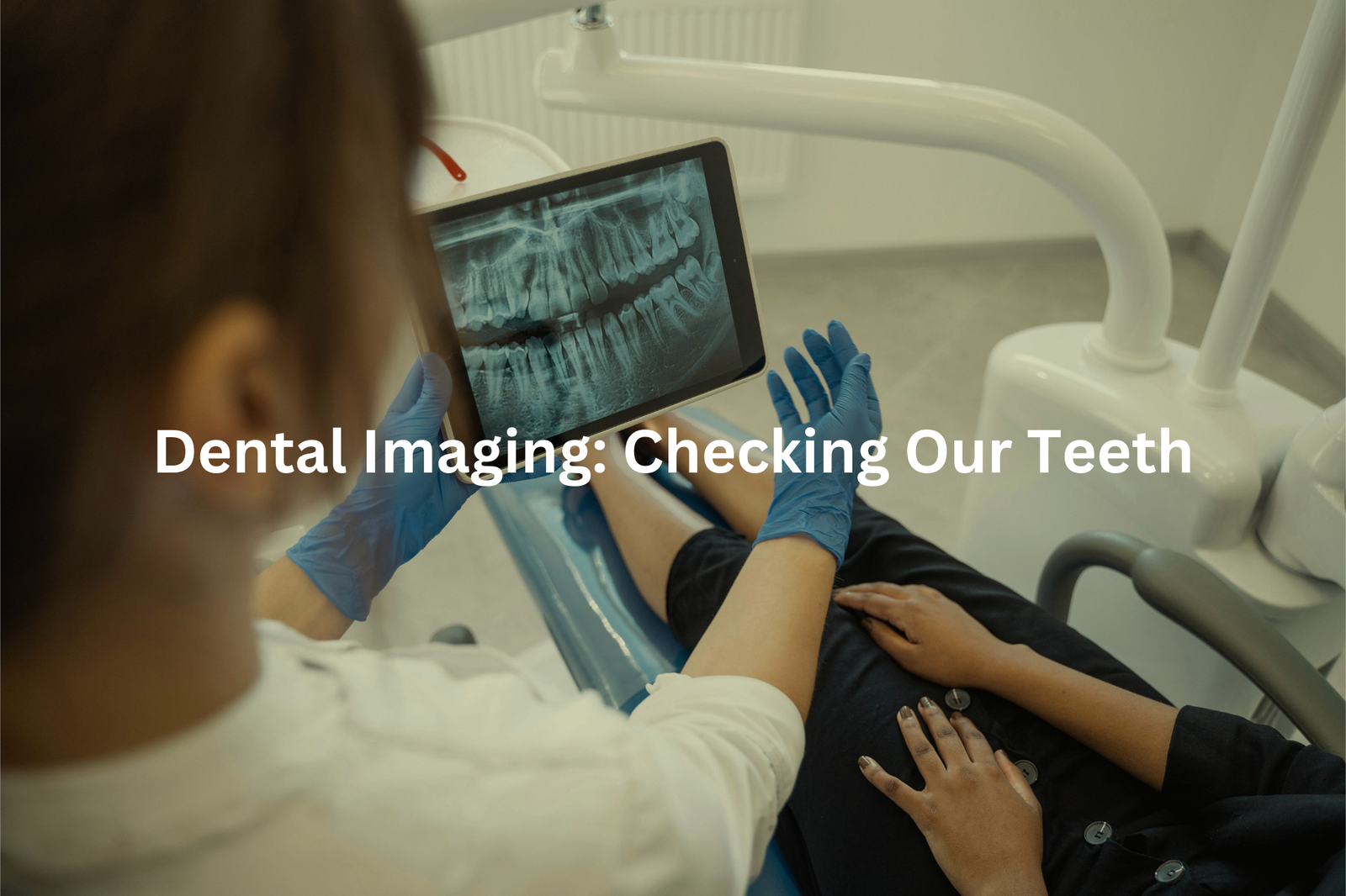
Dentists don’t just clean teeth—they use X-rays to keep them healthy! Those machines that buzz and click during your visit are actually pretty clever. X-rays let dentists see what’s happening under the gums, like cavities or tooth roots that might need attention.
Here’s what X-rays help with:
- Spotting cavities: X-rays can find tiny holes in teeth before they get worse.
- Checking roots: They show if tooth roots are healthy or if there’s trouble.
- Planning treatments: Whether it’s braces or a tooth removal, X-rays help dentists decide the best way forward.
I remember my first dental X-ray. It was quick, just a small machine snapping a picture. I didn’t feel a thing, but the dentist could see everything—cavities, roots, all of it. It felt like magic. So, next time you’re at the dentist, remember: X-rays are like their secret weapon for keeping your teeth strong and healthy.
Foreign Object Detection: Finding Surprises
Sometimes, people swallow things they’re not supposed to, and that’s where X-rays step in. They’re not just for spotting broken bones—they’re like a map for doctors, showing exactly where foreign objects are hiding inside the body.
Here’s what X-rays can do:
- Locate swallowed items: Coins, buttons, or even toys can get stuck. X-rays show exactly where they are.
- Help in emergencies: If someone’s hurt and has shrapnel or other objects in their body, X-rays guide doctors on what to do.
- Spot unexpected things: Swallowed pills or fragments can also show up.
I once read about a boy who swallowed a toy car. The X-ray showed it sitting in his stomach, and doctors removed it safely. So, if something’s gone wrong inside, X-rays are a quick and reliable way to figure it out. They’re small machines, but they make a big difference in saving lives.
Preoperative Assessments: Preparing for Surgery
Before surgery, doctors need to know exactly what’s happening inside the body. That’s where X-rays come in. They give a clear image of bones and joints, helping surgeons plan the operation properly(3). Without them, it’d be like trying to fix something blindfolded—dangerous and unpredictable.
Take knee surgery, for example. An X-ray can show if there’s a fracture, arthritis, or even tiny bone fragments. This helps the surgeon decide the safest and most effective approach. It’s not just about fixing the problem; it’s about doing it right.
Here’s why X-rays are crucial before surgery:
- They reveal hidden injuries or damage.
- They guide the surgeon’s plan step by step.
- They reduce risks by avoiding surprises during the operation.
I once read about a bloke who broke his wrist. His X-ray showed a small, hidden fracture. Without it, the surgery could’ve gone sideways. X-rays make sure surgeons are ready for anything.
Monitoring Treatment Progress: Checking How We’re Doing
Sources: Sky News Australia.
Doctors don’t just use X-rays to find problems; they also use them to check if treatments are working. For example, after a broken bone, X-rays can show if it’s healing properly. If the bone isn’t lining up or connecting, doctors know they might need to adjust the treatment.
The same applies to cancer. After treatments like chemotherapy, X-rays help doctors see if a tumour is shrinking. If it’s not, they can explore other options.
Here’s how X-rays help after treatment:
- Bone healing: X-rays track how a broken bone is mending.
- Tumour size: They show if cancer treatment is reducing tumours.
- Next steps: If results aren’t as expected, doctors adjust plans.
I once read about a patient whose lung tumour shrank after chemo. The X-ray confirmed the progress. Without it, the doctor wouldn’t have known to continue the same treatment. X-rays are like a medical check-in, guiding what happens next.
FAQ
What are X-ray images and how do they work?
X-ray images are a type of medical imaging that uses X-ray beams to create pictures of the body’s internal structures. These X-ray beams pass through the body and are absorbed differently by different tissues, allowing doctors to see bones, organs, and other internal parts. X-ray film or digital X-ray detectors capture these X-ray images, which healthcare providers can then use to diagnose and monitor various medical conditions.
What are the different types of X-ray procedures?
X-ray procedures include standard X-rays, which create images of bones and other internal structures, as well as more specialised X-ray imaging like barium enemas that allow doctors to see the gastrointestinal tract. X-ray technology uses X-ray tubes to generate the X-ray beams, and the amount of radiation exposure is carefully controlled to ensure patient safety during these medical imaging tests.
How much radiation is involved in X-ray exams?
X-ray exams involve exposure to low levels of electromagnetic radiation, which is a type of ionising radiation. The amount of radiation varies depending on the type of X-ray procedure, but healthcare providers carefully monitor the radiation doses to ensure they are as low as possible while still producing high-quality images. Most standard X-rays involve relatively small amounts of radiation, similar to the background radiation we’re exposed to every day.
How do X-rays compare to other medical imaging methods?
X-rays are one of the most common and well-established medical imaging techniques, but they are not the only option. Other imaging methods like magnetic resonance imaging (MRI) and ultrasound use different types of electromagnetic energy beams to create images of the body’s internal structures and tissues. The choice of imaging technique depends on the specific medical issue being evaluated and the information the healthcare provider needs to make an accurate diagnosis.
What are the potential risks of X-ray exposure?
While X-rays are generally very safe, there is a small risk associated with the ionising radiation involved. Exposure to high levels of radiation over a long period of time can potentially increase the risk of cancer or other health problems. However, healthcare providers take extensive precautions to minimise patient radiation exposure during X-ray procedures. The benefits of using X-rays for medical diagnosis and treatment typically far outweigh the small potential risks.
Conclusion
X-rays are super useful in healthcare. They help doctors spot broken bones, look at our lungs, and even peek inside our mouths! X-rays show how we’re healing and can reveal surprises inside our bodies. If you need one, don’t fret! It’s just a quick picture that helps doctors keep you healthy. Always feel free to ask your doctor questions about X-rays and how they can help you stay well. It’s all part of understanding your care.
References
- https://www.qldxray.com.au/services/nuclear-medicine/bone-scan
- https://www.rshq.qld.gov.au/safety-notices/occupational-health/chest-x-ray-image-quality-and-reporting
- https://clinicalexcellence.qld.gov.au/sites/default/files/2018-01/invest-guide.pdf


