

MRI scans use magnets and radio waves to see inside your body. Learn how they work, how to prepare, and stay safe during the process!
MRI scans are vital tools that doctors use to capture images of our bodies. When I first saw one, it was like a fascinating puzzle revealing soft tissues and organs. MRI, which stands for Magnetic Resonance Imaging, uses powerful magnets and radio waves to create these detailed images without any radiation.
Preparing for an MRI usually means removing metal objects and wearing comfortable clothing. Safety is key, as the strong magnets can interact with certain implants. If you’re curious about the procedure or how it feels, keep reading to find out more!
Key Takeaway
- MRI scans use magnets and radio waves to create images.
- Preparation is important for clear results.
- Safety measures help keep patients protected during the scan.
MRI Basics
MRI scans can feel a bit strange at first. They’re big machines that make loud, rhythmic noises—kind of like a jackhammer—but they’re just doing their job. The machine uses powerful magnets and radio waves to create detailed images of the inside of your body. Unlike CT scans, MRIs don’t use X-rays, so there’s no radiation involved, which is a relief for many people.
The machine’s magnet is incredibly strong, much more than a fridge magnet. It interacts with the water molecules in your body, and when they move, they send signals to the machine. These signals are turned into clear, layered images that help doctors see your organs, tissues, and bones.
Sometimes the scan can take up to 45 minutes, and you’ll need to stay still. If you’re claustrophobic, open MRI options might help. It’s loud, sure, but it’s safe, painless, and gives doctors crucial information.
MRI Scan Preparation
Before an MRI, there’s a bit of prep involved, but it’s nothing too tricky(1). You’ll probably need to change into a hospital gown (it’s just easier for the scan) and remove all metal objects like jewellery, glasses, or even hairpins. Metal and MRI machines don’t mix well, so it’s better to leave those behind.
If you’ve got any medical devices—like a pacemaker or an implant—make sure to tell your doctor. They’ll need to know to keep everything safe. Sometimes, you might be asked to drink a contrast dye. It’s a special liquid that helps the machine capture clearer images.
I remember my first MRI prep; I almost walked in with my belt still on. They caught it just in time. It’s easy to forget stuff when you’re nervous. Best tip? Double-check yourself and don’t hesitate to ask questions. It’ll help things go smoothly.
MRI Safety
The first time I had an MRI, I almost forgot to tell them about the metal plate in my leg. Lucky for me, they asked. That’s the thing about MRIs—the magnets are so strong they can pull on metal objects, which can be dangerous. So, it’s really important to let the staff know if you’ve got any metal implants, like joint replacements, screws, or pacemakers.
Here’s what to remember:
- Metal implants: Always tell the staff about any metal in your body. Magnets and metal don’t mix well.
- Claustrophobia: If tight spaces make you nervous, let them know. The MRI machine is like a narrow tunnel, and it can feel a bit much for some people.
- Open MRIs: Ask about open MRI options—they’re less enclosed and might feel more comfortable.
Talk to your doctor about any worries. They’ll help you prepare. It’s all about staying safe and feeling okay during the scan.
MRI for Brain
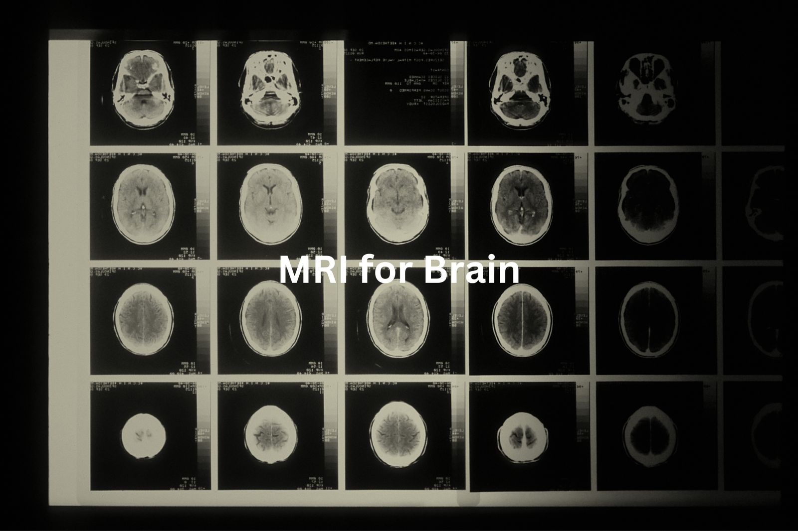
When doctors need to take a closer look at the brain, they often use a brain MRI(2). It’s a type of scan that gives detailed pictures of the brain’s structure. I remember when my uncle had one because his doctor was worried about a tumour. The MRI helped them find it early, which made a big difference in his treatment.
A brain MRI can show things like:
- Tumours: It can spot unusual growths in the brain.
- Blood flow: It shows areas with poor blood flow, which might mean a stroke.
- Brain structure: It reveals the brain’s shape and any changes.
These scans are really clear and help doctors make the best decisions. If there’s a tumour, for example, the MRI shows exactly where it is and how big it is, which helps with surgery or other treatments. If your doctor suggests a brain MRI, it’s just a way to help figure out what’s going on.
MRI for Spine
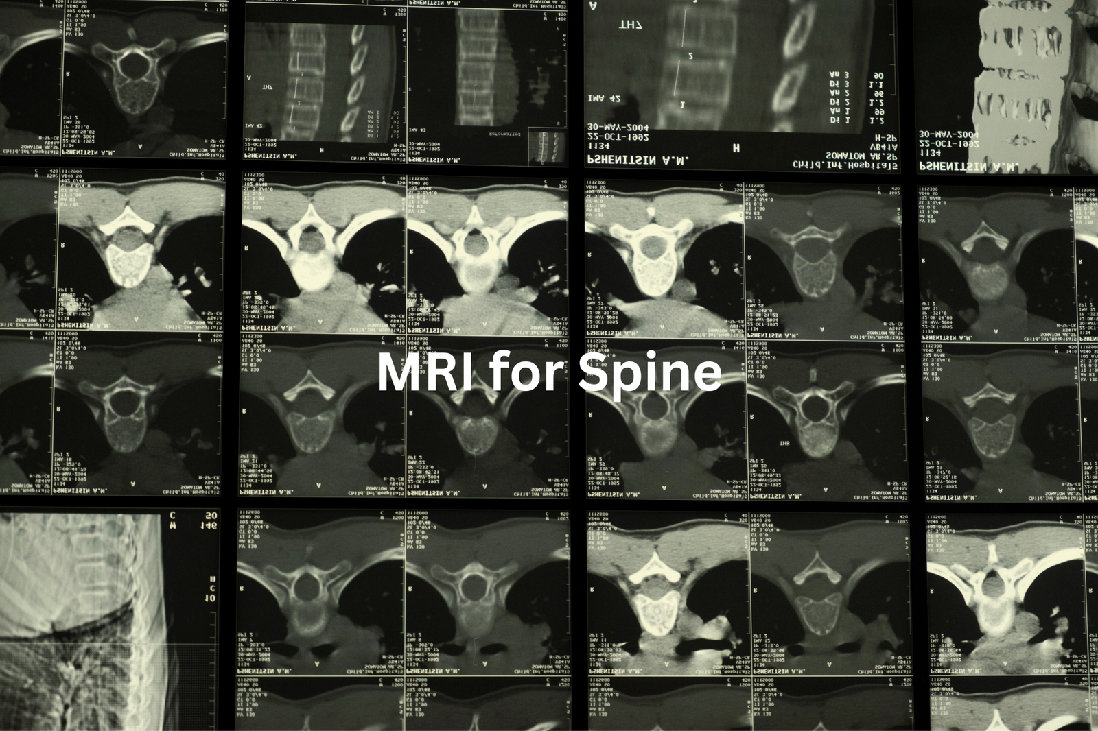
The spine is key to how we move. Without it, we’d be stuck, unable to bend, twist, or even sit properly. When it’s hurt or something’s off, the pain can stop you from doing everyday things. That’s where a spine MRI can help. It gives doctors a detailed look inside.
A spine MRI can(3) find:
- Bulging discs: These can press on nerves and cause pain.
- Injuries: Damage from falls or accidents shows up clearly.
- Soft tissue problems: Muscles and ligaments around the spine can also be checked.
I remember my mate, Sam, had awful back pain. He couldn’t figure out why. Turns out, the MRI showed a bulging disc pressing on a nerve. Without it, the doc might’ve missed the problem. If your back’s been giving you grief, it’s worth asking your doctor if an MRI could help. It’s amazing how much they can see with it.
Contrast MRI
Sometimes, to make MRI scans clearer, doctors use a special dye called a contrast agent(4). It’s like a spotlight, helping certain parts of the body stand out more. This is especially useful for seeing blood vessels, organs, or even small tumours that might be hard to spot otherwise.
Here’s how it works:
- The contrast agent highlights specific areas, making them easier to see.
- It’s injected through a small needle, usually into a vein in your arm.
- It’s generally safe, though some people might have mild allergic reactions, so it’s good to let your doctor know about any allergies.
A mate of mine had chest pain, and the MRI with contrast helped doctors find a tiny blockage in his heart’s blood vessels. Without it, they might’ve missed it. If you’re having an MRI, asking about the contrast agent could be a good idea. It’s not as scary as it sounds and can really help with a more accurate diagnosis.
3T MRI
Sources: Kathleen Vedilago
You might’ve heard of a 3T MRI before. The “T” stands for Tesla, which measures the strength of the magnet inside the MRI machine. A 3T MRI has a stronger magnet than the standard 1.5T MRI found in many hospitals.
Why does this matter?
- Sharper images: The stronger magnet creates clearer pictures, making it easier to spot small details.
- Finding smaller issues: A 3T MRI can detect things like tiny tumours or early signs of disease that might be missed on a 1.5T.
- Advanced imaging: It allows for more detailed scans, helping doctors make better diagnoses.
I remember a friend who had ongoing back pain. A 1.5T MRI didn’t show much, but a 3T scan revealed a bulging disc. It was a game-changer for her treatment. If your doctor suggests a 3T MRI, it’s worth considering. Ask questions so you understand the benefits and feel confident in your choice.
Functional MRI
The brain is like a puzzle, and fMRI (functional MRI) helps us see how the pieces fit together. Unlike a regular MRI, which shows the brain’s structure, fMRI measures blood flow to show activity. When a part of the brain is working, it needs more blood, and fMRI tracks these changes.
It’s used for many things:
- Understanding brain disorders like strokes or Alzheimer’s.
- Researching how we think, feel, and remember.
- Mapping brain activity during tasks like solving puzzles or moving.
I once read about a study where participants solved math problems inside an fMRI machine. Scientists could see which brain areas lit up as they worked. It’s like a peek into the mind. The best part? It’s non-invasive and safe. If you ever need one, it’s just a way for doctors to learn more about how your brain works, helping them help you better.
Open MRI Options
Some people feel really nervous about MRIs. The small tube and loud noises can make it hard to stay calm. That’s why open MRIs are an option. They’re designed to help people who feel anxious in tight spaces.
Here’s how they’re different:
- More space: Open MRIs have a wider opening, so you don’t feel as closed in.
- Same job: They still take images of your body, just like a regular MRI.
- Image quality: Regular MRIs often give sharper pictures because they use stronger magnets. Open MRIs might not be as detailed.
I’ve heard from people who chose open MRIs because they couldn’t handle the regular ones. They said it felt easier to relax and breathe. If you’re worried about an MRI, talk to your doctor. They’ll help you decide which type is better for you. Sometimes, comfort matters more than perfect pictures.
MRI Results
After an MRI, the images don’t go straight to your doctor. First, they’re sent to a radiologist. Radiologists are specialists who study the images carefully. They look for anything unusual—like a broken bone, a tumour, or other issues—and then write a report explaining what they’ve found.
Here’s how it works:
- Radiologist: Reviews the MRI images and looks for problems.
- Report: They write a detailed report about their findings.
- Doctor: Your doctor reads the report and explains the results to you.
Waiting for the report can take a few days, which feels like forever sometimes. I’ve been there—checking my phone every hour, hoping for answers. But the wait is worth it. The report helps doctors understand what’s happening inside your body and plan the next steps, whether that’s more tests or starting treatment. Patience is hard, but the MRI is a big step toward getting answers.
FAQ
What is a CT Scan?
A CT (Computed Tomography) scan uses X-rays to create detailed images of the body’s internal structures. It’s often used to diagnose conditions, check for injuries, or monitor treatment progress. CT scans can provide clear images of bones, organs, and soft tissues, making them a valuable tool in medical diagnosis and treatment planning.
How is an MRI Scan Different from a CT Scan?
While both CT and MRI scans create images of the body, the key difference is that MRI (Magnetic Resonance Imaging) uses powerful magnetic fields and radio waves instead of X-rays. MRI scans can provide more detailed information about soft tissues like the brain, spinal cord, and organs, making them particularly useful for conditions affecting these areas.
What Makes an MRI Scan Safe?
MRI scans are generally considered safe because they don’t use ionising radiation like X-rays. However, the strong magnetic fields used can interact with certain metal objects, so it’s important to inform your healthcare provider about any implants, devices, or metallic objects you may have. Trained professionals ensure the MRI environment is safe and that proper safety protocols are followed.
How Does Quantitative MRI (qMRI) Data Work?
Quantitative MRI (qMRI) involves advanced imaging techniques that provide numerical data about tissue properties, like relaxation times and proton density. This quantitative information can help healthcare providers better understand changes in tissues, potentially leading to earlier disease detection and more accurate monitoring of treatment effects. qMRI data is generated using specialised software and analysis methods.
What are the Benefits of Breast MRI Scans?
Breast MRI scans can be particularly useful for women at high risk of breast cancer, as they can often detect tumours that may be missed by other imaging tests like mammograms. Breast MRI scans provide detailed, high-quality images of the breast tissue, allowing doctors to identify potential issues and guide treatment decisions. This advanced imaging technique is an important tool in the early detection and management of breast cancer.
How Can Foetal MRI Scans Help?
Foetal MRI scans can provide detailed information about a developing baby’s anatomy and potential abnormalities, even in cases where ultrasound results are unclear. These scans use magnetic fields and radio waves to create clear images of the foetus, placenta, and uterus, helping healthcare providers assess foetal development and detect any issues that may require further monitoring or intervention during pregnancy.
What is T2 Mapping in MRI?
T2 mapping is an advanced MRI technique that measures the relaxation time of protons in tissues, known as the T2 relaxation time. This quantitative information can be used to assess changes in tissue properties, such as the water content or degree of degeneration. T2 mapping is particularly useful for evaluating conditions affecting the musculoskeletal system, brain, and other organs.
How Can Cardiac MRI Scans Help?
Cardiac MRI scans use powerful magnetic fields and radio waves to create detailed images of the heart and surrounding blood vessels. These scans can provide valuable information about the heart’s structure and function, helping healthcare providers diagnose and monitor a wide range of cardiac conditions, such as heart disease, valve problems, and congenital heart defects. Cardiac MRI is an important tool in the comprehensive assessment and management of cardiovascular health.
What are the Benefits of Spinal Cord MRI?
MRI scans of the spinal cord can provide healthcare providers with detailed information about the structure and function of this critical part of the nervous system. These scans can help diagnose and monitor a variety of spinal cord conditions, including injuries, tumours, and degenerative diseases. Spinal cord MRI is an essential tool for accurately assessing neurological issues and guiding appropriate treatment and management strategies.
What are the Different Types of MRI Scans?
MRI technology offers a wide range of specialised scan types, each designed to provide specific information about different parts of the body or different tissue properties. Some common MRI scan types include brain MRI, breast MRI, cardiac MRI, and MRI enterography (for the gastrointestinal system). Healthcare providers will select the most appropriate MRI scan based on the patient’s symptoms, medical history, and the suspected condition or area of concern.
Conclusion
MRI scans are important tools that help doctors look inside our bodies without any harmful radiation. They need some preparation, but there are safety measures to keep you safe. Whether it’s for the brain, spine, or other parts of the body, MRI scans give clear pictures that help doctors make the best decisions for your health. So, if you ever need an MRI, think of it like a special camera that helps doctors care for you better!


