
This article explains contrast fluoroscopy, a special imaging technique that helps doctors see inside the body using X-rays and contrast dye.
Have you ever watched a magic show where the magician makes things appear and disappear? Contrast fluoroscopy is a bit like that magic, but instead of tricks, it reveals the mysteries inside our bodies!
This special imaging technique allows doctors to see real-time moving images of different organs and systems. By using a special dye called contrast, they can make certain areas much clearer. Let’s take a closer look at how contrast fluoroscopy works and why it’s so important!
Key Takeaway
- Contrast fluoroscopy uses X-rays and a special dye to help doctors see inside the body.
- It helps diagnose problems in various systems, like the digestive and cardiovascular systems.
- Safety measures are important to protect patients from radiation exposure during the procedure.
What is Contrast Fluoroscopy?
Contrast fluoroscopy feels like watching the inside of the body in motion—a glimpse behind the curtain of organs and systems. Doctors use this X-ray-based imaging to capture real-time video. It’s a key tool for diagnosing issues in various body systems.
In the gastrointestinal system, contrast fluoroscopy helps show how food moves through the stomach or intestines. For the cardiovascular system, it highlights blood vessels, veins, and arteries, making it easier to spot blockages. And in the musculoskeletal system, it shows joint movement or bone structure in precise detail.
This tool doesn’t just take a snapshot; it watches how things work. Its versatility helps identify issues that might not appear on regular still images. While the technique sounds simple, it relies on careful monitoring and radiation safety measures to ensure accuracy and minimise exposure risks. (1)
How Does It Work?
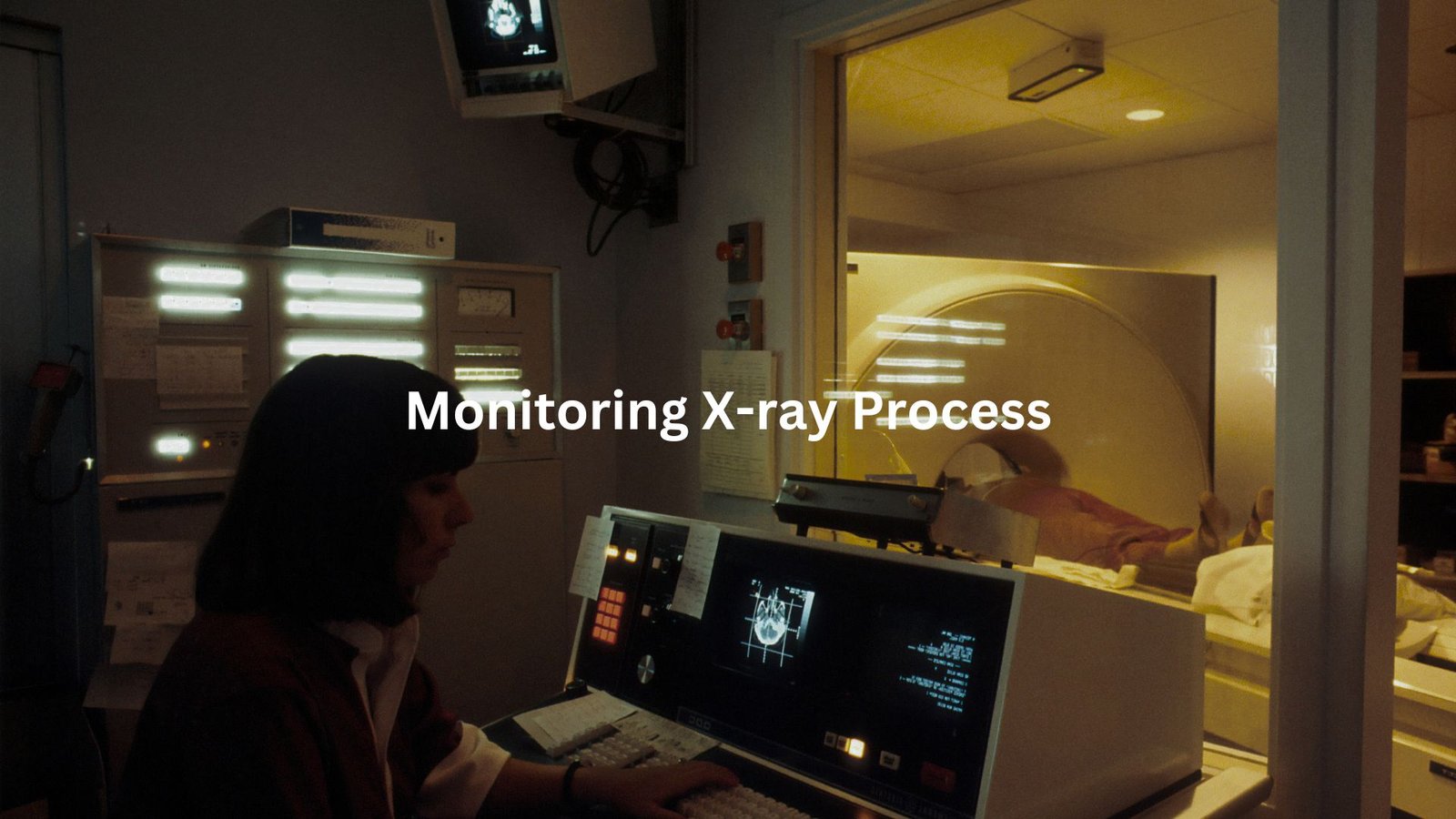
The patient usually lies on a table. The fluoroscope (an X-ray machine used for real-time imaging) hovers over the body.
Doctors first position the patient under the fluoroscope to get the clearest view. Once ready, a contrast dye—often barium or iodine—is injected, swallowed, or delivered through a catheter to the body part being examined. This dye absorbs X-rays, making the internal structures stand out.
As the X-ray beam passes through the body, the fluoroscope sends live images to a monitor. For example, in an ERCP procedure (used to view bile ducts), doctors can see the ducts filling with contrast dye and spot any blockages. It’s like a moving map that helps guide decisions on the spot.
Throughout the process, doctors may adjust kVp or mAs settings to improve image clarity while keeping the radiation dose as low as reasonably achievable (ALARA). The entire procedure depends on precision, and every adjustment matters.
The Importance of Contrast
Credit: Rad Tech Hub By Medical Professionals
Contrast dye is like the highlighter of the medical imaging world—it’s what makes everything pop. Without it, fluoroscopy would be like watching a black-and-white movie with half the picture missing. By adding contrast, doctors get a clearer view of what’s going on inside the body.
Barium is the go-to for digestive system studies. A barium drink coats the stomach and intestines, making them more visible under X-rays. For blood vessels, iodine is used. It brightens up arteries and veins so blockages or narrowing are easier to see. Gastrografin is another option when barium isn’t suitable, especially for patients at risk of perforations.
Contrast helps reveal issues like blockages, strictures (narrowing of the digestive tract), or abnormal blood flow. While it can sometimes cause mild side effects, such as nausea, the benefits of spotting problems early far outweigh the risks. Proper use of contrast is essential for accurate diagnosis and treatment. (2)
Types of Procedures
Contrast fluoroscopy is used across many types of procedures, each tailored to spot different issues. The barium swallow, for example, is simple but powerful. A patient drinks a barium solution, and doctors watch how it travels through the oesophagus, looking for things like reflux or narrowing.
For cardiac angiography, doctors inject iodine contrast into a catheter. It flows through the arteries, showing any blockages or irregularities in blood flow to the heart. This can help guide decisions about treatments like stents.
Another common procedure is the barium enema. In this test, barium is introduced through the rectum to visualise the large intestine. It’s particularly useful for spotting issues like polyps or diverticulitis.
Each of these procedures relies on careful positioning and timing to minimise radiation exposure while getting the clearest image possible. And all are part of a larger safety-first approach to medical imaging.
Safety Considerations
Safety Precautions in Contrast Fluoroscopy
Safety isn’t just a checkbox during contrast fluoroscopy—it’s the backbone of the entire process. While the images help save lives, X-rays come with risks, like radiation exposure. The challenge is keeping those risks as low as reasonably achievable (the ALARA principle) while still getting a clear image.
Medical staff often wear lead aprons. You’ll see these heavy, awkward shields draped over their torsos. They might not look comfortable, but they work. Lead blocks scatter radiation, reducing exposure to sensitive organs. Thyroid shields protect the neck area, and lead glasses can help shield eyes from radiation that could lead to cataracts over time.
Dose monitoring is another layer of protection. Modern fluoroscopy units have built-in dosimeters that track how much radiation is used in real-time. This monitoring ensures the absorbed dose stays below harmful levels for both patients and staff.
Time and distance play a big role too. Shorter exposure times mean less radiation. It’s that simple. And by positioning themselves further from the X-ray beam, staff can cut their exposure even more. (3)
One final safeguard: regular safety checks on equipment to catch any radiation leaks or malfunctions. Fluoroscopy is a powerful tool, but it’s most effective when safety is the first thought, not the last.
FAQ
What is contrast fluoroscopy, and how does it work?
Contrast fluoroscopy uses a contrast dye and an X-ray machine to create real-time images. It’s a powerful tool for diagnosing issues in various systems, like the digestive system or cardiovascular system. The dye—often barium sulfate or iodine—helps highlight tissues, blood vessels, and organs.
A radiologist monitors the images for abnormalities, such as strictures or blockages. The image intensifier improves clarity, showing even tiny details in real-time. This technique helps doctors evaluate conditions and guide interventions with precision.
Why is contrast dye used in fluoroscopy?
Contrast dye is essential because it enhances visibility during fluoroscopy. Positive contrast agents (like barium sulfate and iodine) make tissues or structures appear bright on X-rays, while negative contrast agents (like air) create darker areas. This difference helps spot abnormalities, such as lesions, strictures, or reflux. Double contrast studies use both types to increase detail. The contrast also allows real-time evaluation of motility and organ function, making diagnosis more accurate.
What types of procedures use contrast fluoroscopy?
Contrast fluoroscopy is used in many procedures, including:
- Barium swallow and enema: Examines the digestive system for blockages, strictures, or perforation.
- Cardiac angiography: Checks heart arteries for blockages or narrowing.
- Digital subtraction angiography (DSA): Enhances blood vessel images using a mask image.
These procedures help doctors monitor and guide interventions with precision.
How is safety managed during contrast fluoroscopy?
Safety is managed by limiting radiation exposure and using shielding devices like lead aprons and thyroid shields. Radiologists monitor the X-ray dose to reduce risks to the kidneys, liver, and other organs. Regular safety checks on the C-arm and other imaging equipment ensure everything works correctly. For patients, using only the necessary dose helps reduce potential long-term risks.
What is digital subtraction angiography (DSA), and how is it different?
DSA is a form of contrast fluoroscopy used in interventional radiology. It captures two images: a mask image (taken before the contrast injection) and a contrast medium-enhanced image. The mask is subtracted from the contrast image, highlighting blood vessels without showing surrounding tissues. This method is commonly used to evaluate cardiovascular abnormalities or guide catheter placement during interventions.
Can contrast fluoroscopy detect abnormalities in the digestive system?
Yes, contrast fluoroscopy is commonly used to diagnose digestive system issues. Barium swallow or enema procedures can detect strictures, blockages, reflux, or aspiration. Gastrografin, another contrast medium, may be used for patients at risk of perforation or aspiration. Real-time imaging helps monitor motility, ensuring the digestive system functions properly.
What role does contrast fluoroscopy play in interventional radiology?
In interventional radiology, contrast fluoroscopy guides minimally invasive procedures, such as catheter placement or foreign object removal. It provides real-time imaging of blood vessels, helping doctors target treatment areas accurately. This technique is critical for coronary and orthopedic interventions, reducing surgical risks by improving precision.
What are the risks of contrast fluoroscopy, and how are they managed?
Contrast fluoroscopy involves some risks, such as radiation exposure and potential allergic reactions to contrast dye. Radiologists manage radiation exposure with shielding and dose monitoring. They also evaluate kidney function beforehand, especially when using iodine-based contrast. For those with allergies, alternative dyes or pre-treatment options may be considered.
Conclusion
Contrast fluoroscopy is a powerful imaging technique that allows doctors to see moving images inside the body using X-rays and special dye. This method is essential for diagnosing conditions in various systems, such as the gastrointestinal and cardiovascular systems. However, safety measures are crucial to protect patients and staff from radiation exposure.
By using contrast effectively and following safety guidelines, doctors can uncover important information that helps guide treatment decisions. So, the next time you hear about contrast fluoroscopy, remember it’s all about making the invisible visible!
References
- https://pubmed.ncbi.nlm.nih.gov/34283448/
- https://www.uclh.nhs.uk/patients-and-visitors/patient-information-pages/contrast-agents-x-ray-fluoroscopy-ct-and-angiography-examinations
- https://my.clevelandclinic.org/health/diagnostics/21992-fluoroscopy


