
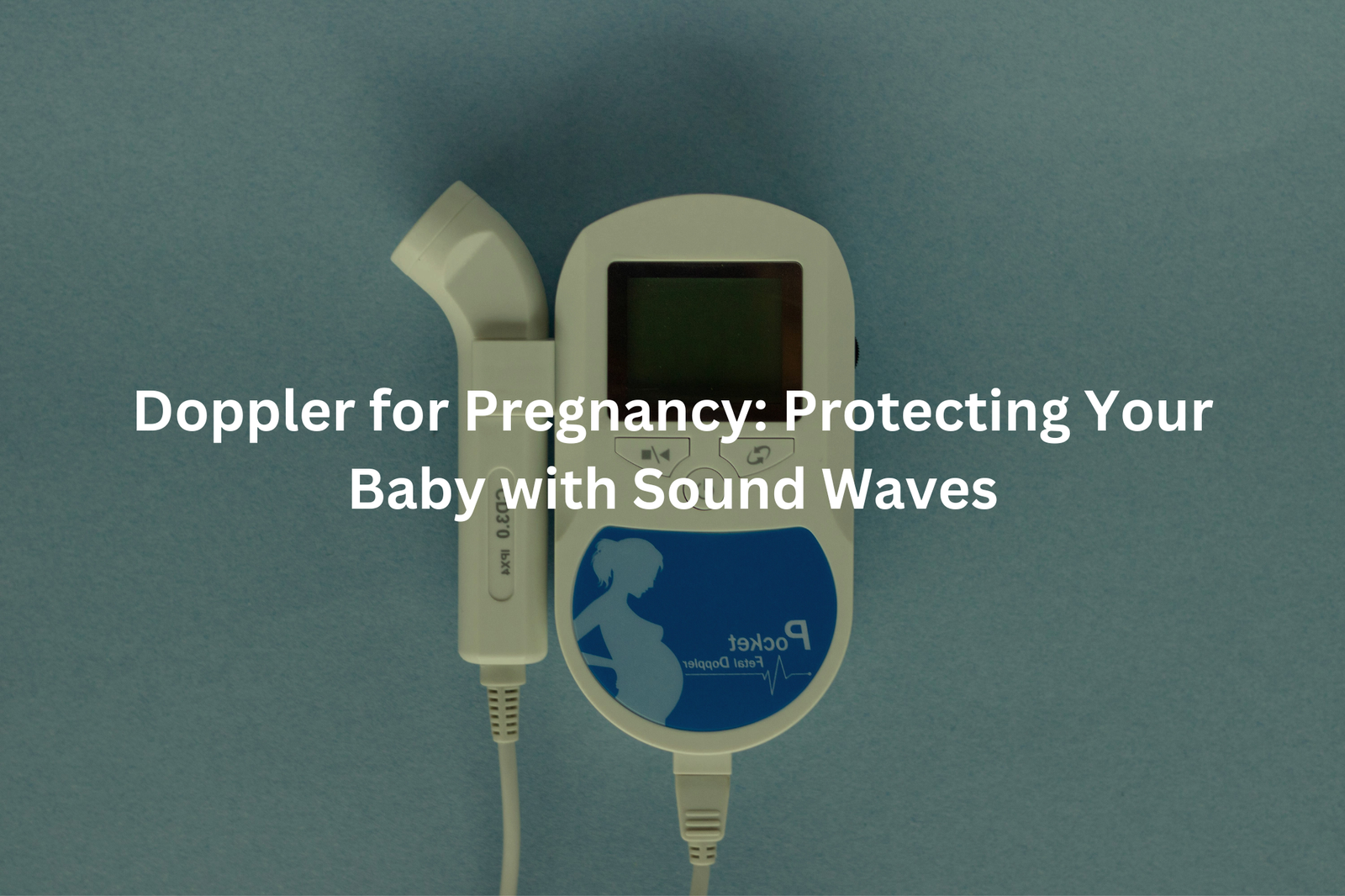
Doppler for pregnancy uses sound waves to check blood flow and growth. It’s an easy, non-invasive way to keep both mum and baby safe during pregnancy!
Doppler ultrasound helps doctors check on babies before they’re born. This medical tool sends sound waves through the mum’s belly (operating at frequencies between 2-18 MHz) to watch how blood moves in the baby and placenta.The machine shows blood flow patterns on a screen, letting doctors spot any problems early.
They can check the baby’s growth, heart, and blood vessels. Most mums get these scans during routine check-ups to make sure everything’s going well. If you’re curious about how ultrasound works and what it can do, you might want to read more about it. It’s a fascinating topic!
Key Takeaways
- Doppler ultrasound checks blood flow in babies and the placenta.
- It helps doctors find out if there are any problems early.
- Using Doppler can make birth safer for both mums and babies.
Understanding Doppler Ultrasound
Doppler ultrasound serves as a crucial monitoring tool during pregnancy, using sound waves to track blood flow patterns between mother and baby. The non-invasive test, commonly performed after 20 weeks of pregnancy, measures blood movement through essential vessels.
Medical professionals focus on two key areas during the scan(1):
- Umbilical artery: Shows blood flow from mum to bub
- Middle cerebral artery: Monitors blood supply to the baby’s brain
The procedure takes about 15 minutes (give or take a few), with the sonographer applying gel to the pregnant belly before moving a small handheld device across the skin. No needles needed, just gentle pressure.
This scan becomes particularly valuable for high-risk pregnancies, such as those involving:
- Gestational diabetes
- High blood pressure
- Multiple pregnancies
- Previous pregnancy complications
The results help doctors spot potential issues early on, letting them adjust care plans when needed. Regular monitoring through Doppler ultrasound helps ensure both mum and bub stay healthy throughout pregnancy.
Why is Doppler Important for High-Risk Pregnancies?
Sources: yes OR no.
High-risk pregnancies need careful watching, and Doppler ultrasound stands as a key medical tool(2). This technology spots potential issues before they turn serious, giving doctors a chance to act quickly.
The scan focuses on three main blood vessels:
- The umbilical artery (carrying oxygen to the baby)
- The middle cerebral artery (showing brain blood flow)
- The placental vessels (checking nutrient delivery)
Medical studies show that Doppler ultrasound reduces baby deaths by 29% in high-risk cases. The test helps doctors decide if they need to deliver early or plan a C-section.
For example, if blood flow looks weak through the umbilical cord, doctors might choose to deliver the baby sooner. The scan takes about 30 minutes (using special gel and a handheld device), and it doesn’t hurt at all.
Doctors suggest getting these scans every few weeks during high-risk pregnancies. They’re quick, safe, and might spot problems before they become dangerous.
Doppler Use in Low-Risk Pregnancies
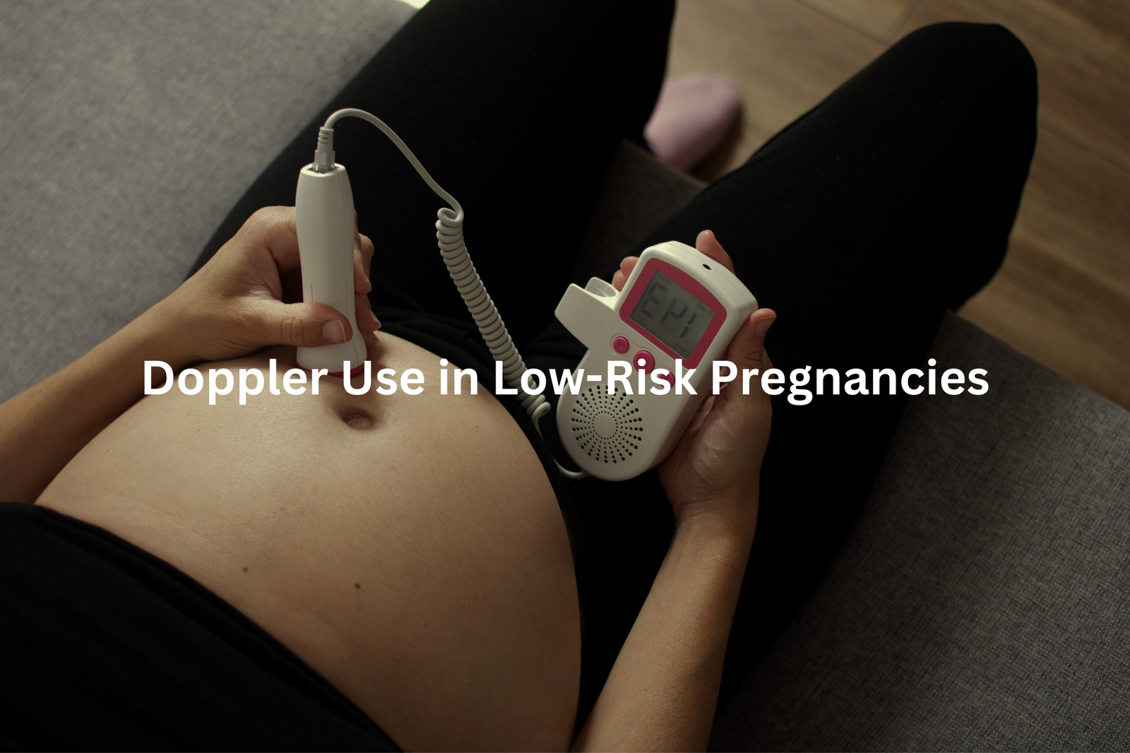
Doppler ultrasound scans serve as valuable tools in pregnancy monitoring, going beyond their traditional use in high-risk cases. These scans measure blood flow in three key areas (the umbilical artery, middle cerebral artery, and placental vessels) to spot potential issues before they become serious.
Medical professionals use this technology to check(3):
- Blood flow between mum and bub through the umbilical cord
- Oxygen delivery to the baby’s brain
- How well the placenta works
The debate around routine Doppler use continues in Australian medical circles. While some doctors recommend these scans for all pregnancies, others suggest limiting them to high-risk cases. Current research shows promising results for early detection of complications, even in seemingly normal pregnancies.
For expectant mums considering this option, a chat with their healthcare provider can help determine if Doppler monitoring fits their pregnancy journey. The scan takes about 20 minutes and doesn’t cause any discomfort to mum or bub.
How is a Doppler Ultrasound Done?
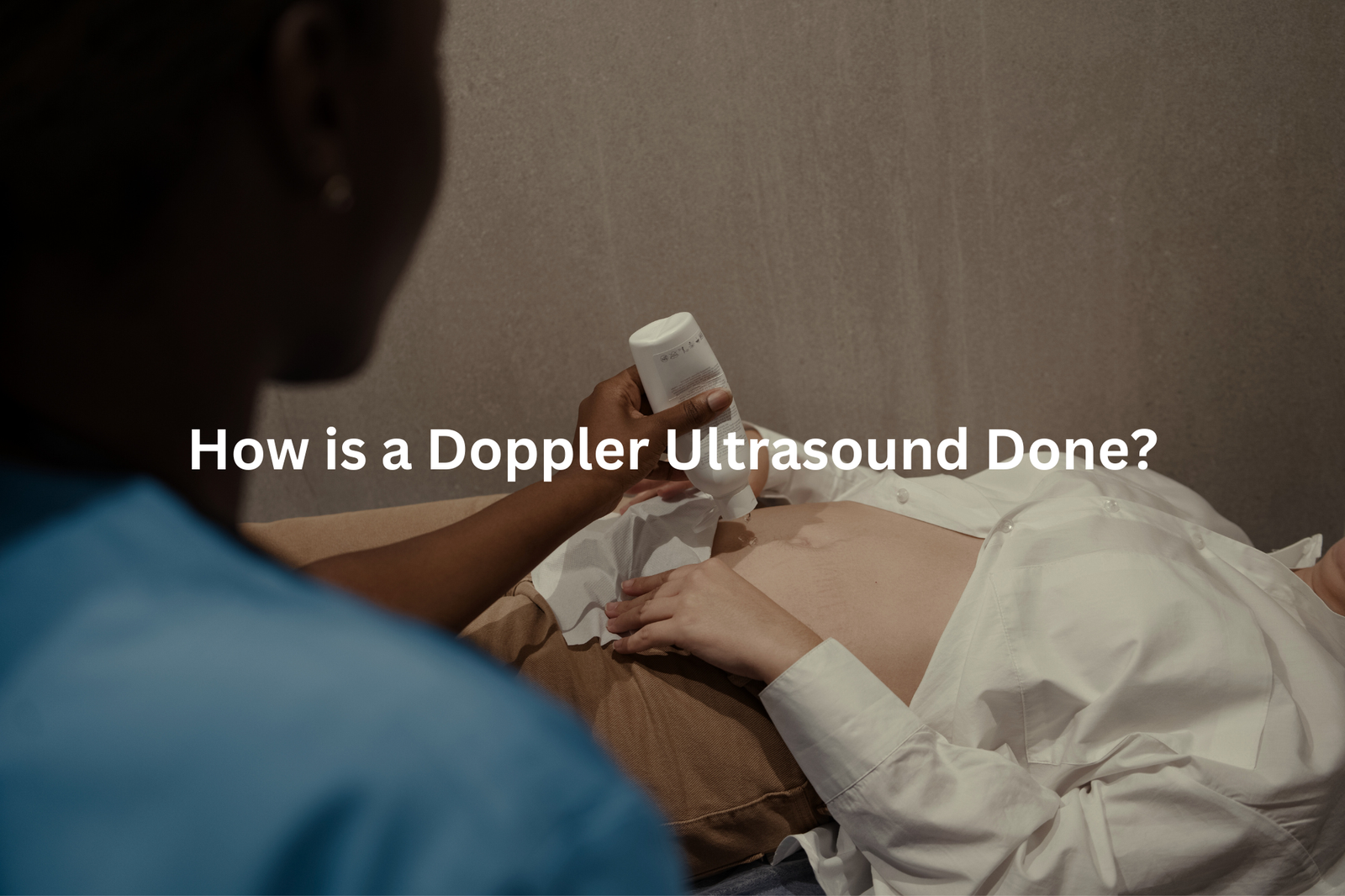
A Doppler ultrasound scan helps doctors check blood flow in unborn babies. The procedure takes about 30 minutes and uses sound waves to create detailed images (operating at frequencies between 2-18 MHz).
The process starts when a patient lies on an examination table. Medical staff applies a water-based gel to the abdomen, which acts as a conductor for the ultrasound waves. A handheld device, called a transducer, moves across the gel-covered area to capture images.
During the scan, the transducer measures:
- Blood flow through the umbilical cord
- Circulation in the baby’s brain
- Movement through major blood vessels
The gel might feel cool on the skin, but the procedure causes no discomfort. Patients don’t need special preparation before the scan, though wearing loose-fitting clothes helps. The ultrasound waves pose no risk to mother or baby, making this test a safe way to monitor pregnancy health.
Medical staff can explain the results right after the scan, showing the blood flow patterns on a nearby screen.
Outcomes and Evidence
Doppler ultrasound stands as a vital monitoring tool for pregnant women, particularly those with high-risk pregnancies. This medical technology uses sound waves (operating at frequencies between 2-18 MHz) to check blood flow between the mother and baby.
Research points to three main benefits of Doppler ultrasound:
- Reduces unnecessary medical procedures, letting more women experience natural births when safe
- Helps track proper foetal growth and development, leading to better birth weights
- Lowers perinatal death rates through early detection of potential problems
Medical professionals use this technology to examine blood flow in the umbilical cord and major blood vessels. The process takes about 20 minutes, doesn’t hurt, and needs no special preparation.
For high-risk pregnancies, doctors might suggest Doppler scans every two to four weeks. Regular monitoring helps spot issues early, giving doctors time to adjust care plans. Pregnant women should chat with their healthcare provider about whether Doppler monitoring suits their situation.
FAQ
What is the role of low-risk and high-risk Doppler in pregnancy?
Doppler ultrasound is a valuable tool for monitoring the health of both low-risk and high-risk pregnancies. For low-risk pregnancies, Doppler can help check on the baby’s growth and development. For high-risk pregnancies, Doppler can detect potential issues with the baby’s blood flow, heart rate, and other vital signs, allowing healthcare providers to intervene early if needed.
How do Doppler artery PI and RI values indicate fetal well-being?
The pulsatility index (PI) and resistance index (RI) measured in the baby’s arteries using Doppler can provide insights into the health of the placenta and fetal circulation. Abnormal PI and RI values may signal issues like fetal growth restriction or placental problems, allowing doctors to closely monitor the pregnancy.
What information can Doppler heart rate monitoring provide during pregnancy?
Doppler can be used to continuously monitor the baby’s heart rate throughout pregnancy. This helps detect any concerning changes in the heart rate that could indicate fetal distress, allowing for prompt medical intervention if needed. The heart rate data can also provide clues about the baby’s overall wellbeing.
How do Doppler studies on the uterine, umbilical, and fetal arteries help assess risk factors?
Doppler studies on the blood flow in the mother’s uterine arteries, the baby’s umbilical cord, and the baby’s own arteries can reveal important information about potential risk factors. Abnormal flow patterns may signal issues like placental dysfunction, fetal growth problems, or other complications that require closer monitoring or treatment.
What role does Doppler play in evaluating fetal growth and birth outcomes?
Doppler ultrasound can be used to assess fetal growth throughout pregnancy. Measurements of blood flow in the umbilical cord and fetal arteries, as well as the baby’s size, can help identify potential growth issues early on. This information can then be used to predict birth outcomes and adjust prenatal care as needed.
How can Doppler help detect fetal distress and prevent complications?
By closely monitoring the baby’s blood flow and heart rate using Doppler, healthcare providers can detect signs of fetal distress, such as reduced blood flow or irregular heart patterns. This allows them to intervene promptly to prevent serious complications and ensure the best possible outcome for the mother and baby.
What current research on Doppler in pregnancy is available?
Recent studies using large sample sizes and meta-analyses have examined the use of Doppler in a variety of pregnancy scenarios. This research has helped clarify the role of Doppler in assessing risk factors, detecting issues like fetal growth restriction, and predicting birth outcomes – all of which can improve prenatal care and support healthy pregnancies.
Conclusion
Doppler ultrasound stands as a key medical tool in pregnancy monitoring. This device (using sound waves at 3-10 MHz) watches blood flow patterns in both mother and baby, making it easier to spot problems before they get worse. For high-risk pregnancies, doctors use it to check the baby’s blood vessels and heart function.
Research shows better outcomes with regular monitoring – babies tend to have better birth weights, and there’s less chance of complications. While doctors still study its role in low-risk cases, the technology helps create safer pregnancies through early detection.
References
- https://envisionmi.com.au/us-doppler/
- https://learn.stillbirthcre.org.au/wp-content/uploads/2023/08/CRE_Home-Doppler-statement.pdf
- https://www.pregnancyparenting.org.au/pregnancy/ultrasound


