
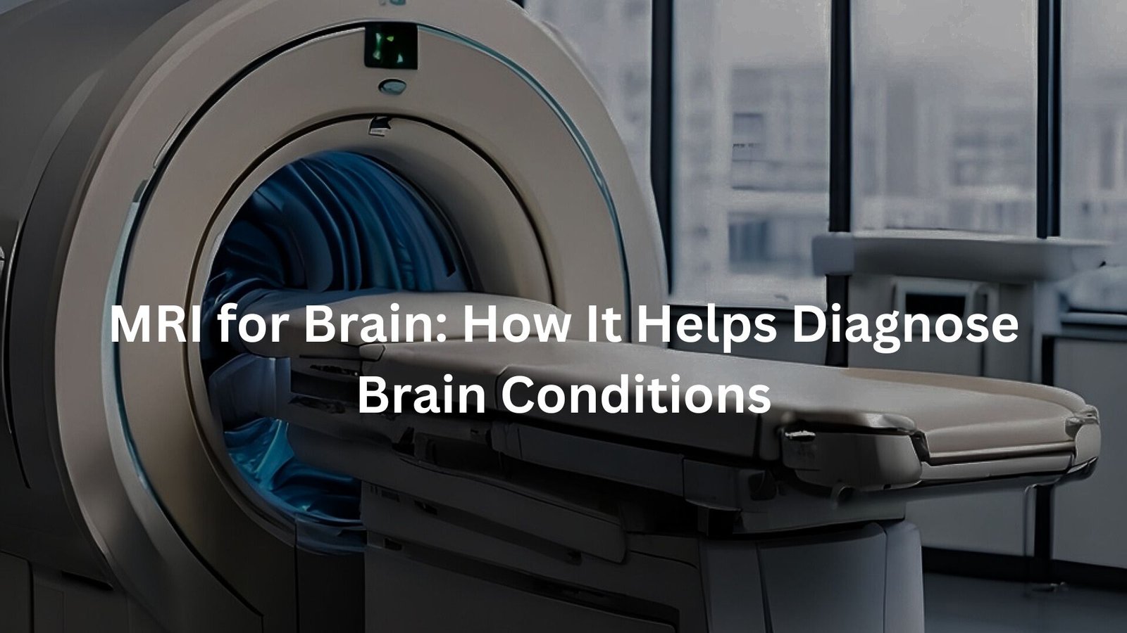
Learn how MRI scans can safely identify brain conditions, from tumours to strokes, with high accuracy.
MRI for brain imaging is a vital diagnostic tool used to identify various brain conditions. By using strong magnetic fields and radio waves, MRIs produce clear, detailed images that assist doctors in diagnosing everything from brain tumours to strokes.
This non-invasive procedure is essential for assessing neurological health and planning treatments or surgeries, offering a safer alternative to other imaging methods.
Key Takeaway
- MRI uses strong magnetic fields and radio waves to create detailed brain images.
- It is essential for diagnosing conditions like brain tumours, strokes, and injuries.
- MRI is non-invasive, offering a safer, radiation-free option compared to other imaging tests.
How MRI for Brain Imaging Works

MRI (Magnetic Resonance Imaging) is a breakthrough in medical imaging, using powerful magnets and radio waves to capture detailed images of the brain.
When placed inside the machine, protons in the body align with the magnetic field, and radiofrequency pulses disturb this alignment, causing protons to release signals. These signals are then transformed into high-resolution brain images.
Key details:
- MRI provides unmatched detail of brain structures.
- It helps doctors diagnose conditions with precision.
Benefits of MRI for Brain Imaging
What makes MRI stand out? For one, there’s no ionising radiation, unlike X-rays or CT scans. This makes it safer for repeated use, especially for patients needing regular monitoring, such as those with chronic conditions or at risk of strokes or brain tumours.
Key advantages:
- MRI offers superior contrast for soft tissues, allowing doctors to see brain structures in fine detail.
- It’s crucial for detecting vascular issues like aneurysms or strokes, offering clearer images of blood flow. (1)
MRI Applications in Diagnosing Brain Conditions
MRI is a vital tool in diagnosing various brain conditions. It’s particularly effective at detecting brain tumours, whether primary or metastatic. Tumours appear as distinct areas in the brain, and MRI can accurately pinpoint their location, size, and characteristics.
MRI is also crucial for stroke detection. It can differentiate between:
- Ischaemic strokes (blocked blood flow)
- Haemorrhagic strokes (bleeding)
For demyelinating diseases like multiple sclerosis (MS), MRI reveals damage to myelin, the protective nerve sheath. Additionally, MRI helps assess brain trauma, determining the extent of damage after accidents or infections and guiding treatment decisions.
MRI for Brain Surgery Planning
MRI is essential in brain surgery planning, offering a detailed, high-definition view of the brain’s anatomy. This information helps surgeons understand the brain’s shape, structure, and any abnormalities, which is critical for a successful operation.
- Pinpoints tumour location: MRI identifies the precise location of tumours or other issues.
- Assess proximity to critical areas: It helps determine how close a tumour is to vital brain regions like the motor cortex or language centres.
With these insights, surgeons can plan their approach with greater precision, reducing risks and improving surgical outcomes. (2)
Safety Guidelines for MRI Brain Scans
Safety is a top priority during MRI scans. Before entering the machine, patients undergo a thorough screening to ensure there are no contraindications, such as metal implants or pacemakers, which can be dangerous in the magnetic field. It’s also important to remove all metal objects, including jewellery, hearing aids, and dental work.
- Screening for safety: Ensure no metal implants or pacemakers are present.
- Claustrophobia concerns: MRI machines can feel enclosed, and noise can be overwhelming.
To help with anxiety, many facilities provide earplugs, sedation, or calming techniques, and ear protection reduces the loud noise significantly.
Common Side Effects of MRI Brain Scans
While MRIs are generally safe, it’s important to be aware of potential side effects. One common issue is an allergic reaction to the contrast agents used in some procedures. These agents help highlight areas of the brain, but they can occasionally cause a rash or itching.
- Possible allergic reactions: Rash or itching due to contrast agents.
- Metallic taste: Some patients may experience a temporary metallic taste after receiving contrast.
- Nephrogenic systemic fibrosis (NSF): A rare condition triggered by contrast agents in patients with kidney problems.
Thankfully, these reactions are uncommon, and most side effects are temporary.
What to Expect During an MRI Brain Scan
Getting an MRI is a simple process, though it might feel a bit intimidating if it’s your first time. You’ll lie down on a bed, and a coil will be placed around your head to help focus on your brain’s details. The scan itself can last from 20 minutes to an hour, depending on the required images.
- Stay still: It’s essential to remain as still as possible to ensure clear, sharp images.
- Noise: The machine will make buzzing or thumping noises, but ear protection is usually provided to reduce the sound.
The process is smooth and painless, with staff on hand to guide you.
Tips for Claustrophobic Patients
Credits: Siemens Healthineers
Claustrophobia can be a common worry for people heading into an MRI scan. The enclosed, narrow space can feel a bit overwhelming, but there are options to make the experience more comfortable.
- Open MRI: Some clinics offer open MRIs, which provide a less confined space, making it easier to feel more at ease. These machines allow you to lie in a more open position, which can help alleviate feelings of anxiety.
- Sedation: If an open MRI isn’t available or you’re still feeling anxious, sedation might be an option. This can help you relax and make the process smoother.
- Pre-scan discussion: Always speak with your doctor beforehand if you’re feeling nervous. They can guide you on available options and ensure that all arrangements are made to keep you comfortable and calm during the scan.
Rest assured, there are ways to manage the experience and make it as stress-free as possible.
Conclusion
In conclusion, MRI is a vital tool for brain imaging, offering clear, detailed views without the risks of radiation. It helps doctors diagnose a wide range of conditions, from tumours to strokes, and plays a key role in surgery planning.
With its non-invasive nature and ability to detect fine details, MRI continues to be an indispensable part of modern medicine, providing critical insights for patient care and improving outcomes in brain health.
FAQ
What is Magnetic Resonance Imaging (MRI) for the brain?
Magnetic resonance imaging, or MRI, is a medical test that uses powerful magnets and radio waves to create detailed images of the brain. The MRI scan helps doctors see soft tissues, like the brain structures, blood vessels, and even the internal structures of the brain. MRI is often used to detect brain tumours, brain injury, and other medical conditions that affect the brain tissue.
How do MRI scans work?
During an MRI scan, strong magnets align the protons in your body, and then pulses of radio waves are sent into your body. This causes the protons to emit radio signals, which are captured to form detailed images. The high-resolution images allow doctors to look at the brain in fine detail, showing everything from the frontal lobes to the pituitary gland and brain stem.
Are there any risks with MRI scans?
Generally, MRI scans are considered very safe, as they don’t use harmful ionising radiation like X-rays or CT scans. However, patients with certain medical conditions, like kidney disease or implanted medical devices, should inform their doctor before the scan. Metallic items, such as metal zippers, body piercings, and electronic devices, must be removed as they can interfere with the powerful magnets used in MRI. If you have any concerns, discuss them with your healthcare provider.
Can MRI scans help detect brain tumours?
Yes, MRI scans are one of the most effective imaging methods for detecting brain tumours. These scans provide high-resolution images of brain tissue and help doctors pinpoint the location, size, and type of tumour. MRI can also help doctors determine if a brain tumour is affecting blood flow or nearby blood vessels.
What if I have metal in my body, like an artificial joint?
MRI scans use strong magnets, so patients with certain metal items, such as artificial joints or metal hair accessories, may face risks. Foreign body objects like metal fragments can be dangerous, as they might move or heat up in the magnetic field. Be sure to inform your doctor about any medical devices, like nerve stimulators or pacemakers, that may be inside your body.
Can MRI help with brain surgery planning?
Yes, MRI is a key tool for planning brain surgery. Before surgery, detailed images of the brain are taken to understand the location of the problem, whether it’s a brain aneurysm, brain damage, or brain injury. This helps surgeons plan the procedure by looking at the brain’s internal structures and ensuring a safe and precise approach to the surgery.
Are there any side effects from MRI scans?
Most people don’t experience side effects from an MRI scan. However, in some cases, patients may have allergic reactions to the contrast agents used in the MRI, leading to symptoms like a rash or itching. Additionally, some patients report a metallic taste in their mouth after receiving a contrast agent, though this typically fades quickly. If you have concerns about allergic reactions, speak with your doctor before your scan.
How long does an MRI scan take?
An MRI scan for the brain typically lasts between 20 minutes and an hour. It’s important to stay still during the scan so that the MRI can capture clear and detailed images. The machine may make loud thumping or buzzing noises, so earplugs are often provided. If you’re claustrophobic or feel anxious, your doctor may offer calming techniques, like anti-anxiety medication, to help you relax.
Is MRI useful for diagnosing brain conditions like strokes?
MRI is excellent for diagnosing brain conditions such as strokes. It can detect ischaemic strokes caused by blocked blood flow, or haemorrhagic strokes caused by bleeding. MRI also provides high-quality images of the blood supply and blood vessels in the brain, making it easier for doctors to assess the extent of damage caused by a stroke.
What should I avoid before an MRI scan?
Before an MRI scan, avoid wearing any metallic items, such as credit cards, underwire bras, or metallic hair accessories. These objects can interfere with the MRI machine’s strong magnets. It’s also important to remove any electronic devices, like mobile phones or electronic items, before entering the MRI room. Be sure to inform your doctor about any medical devices or implants, such as nerve stimulators or pacemakers, you might have.
References
- https://www.racgp.org.au/afp/2013/november/mri-brain-imaging
- https://www.melbourneradiology.com.au/diagnostic-imaging/mri-scan-brain/


