
Explore the benefits of X-ray scans in diagnosing injuries, detecting tumours, and ensuring safety in medical imaging.
X-ray scans are essential in modern medicine, providing critical insights into the body’s internal structures. By using electromagnetic radiation, they create detailed images that help healthcare professionals detect fractures, tumours, and other health conditions.
While X-rays come with some radiation exposure, the advantages of early diagnosis often outweigh the risks, making them a crucial part of medical care.
Key Takeaways
- Types of X-ray Scans: Standard X-rays, CT scans, and DEXA provide targeted insights for various health conditions.
- Radiation Safety: X-ray scans use controlled radiation to minimise risks, with safety measures like lead aprons.
- Applications in Cancer & Orthopedics: X-rays are key in diagnosing fractures and detecting tumours, playing a role in cancer monitoring.
What are X-ray Scans?

X-ray scans are a vital part of modern healthcare. They’re a form of electromagnetic radiation that can pass through the body, helping doctors see the inside of a patient’s body without needing to make an incision. X-rays are absorbed by different types of tissues in varying degrees, which is what creates the images that doctors rely on to make diagnoses.
An X-ray machine works by emitting a controlled amount of radiation that moves through the body, hitting a detector on the other side. The detector captures the X-rays that pass through and forms an image. Denser structures, like bones, absorb more radiation, which results in a white image, while softer tissues, like muscles, absorb less, appearing darker on the image.
Components of an X-ray Machine:
- X-ray Source: The part of the machine that generates the X-rays.
- Tubes: Directs the X-rays through the body.
- Detectors: Capture the X-rays that pass through and create the images.
In this way, X-rays help doctors “look” inside the body, providing a clear view of bones, organs, and even blood vessels. (1)
Types of X-ray Scans
X-ray technology has evolved, and now there are several different types of scans, each suited to different medical needs.
Standard X-rays:
These are the most common and are used for general imaging. They’re ideal for diagnosing fractures, detecting pneumonia, and examining the overall health of the bones.
Fluoroscopy:
This type of scan provides real-time imaging. It’s often used for procedures like a barium swallow (used to examine the digestive system) or to guide a doctor through a surgery or other medical procedure.
CT Scans:
A computed tomography (CT) scan is essentially a series of X-rays taken from different angles. These images are then combined to form a detailed 3D image of the area being examined. CT scans are used for everything from detecting cancer to identifying internal injuries and problems with organs.
Dual-Energy X-ray Absorptiometry (DEXA):
DEXA scans are specifically used to measure bone density. This type of X-ray can help identify conditions like osteoporosis, a disease where bones become fragile and break easily. (2)
Key Applications of X-ray Scans
X-ray scans have a range of uses in medicine, and they’re incredibly important in helping doctors make accurate diagnoses. Here’s how they’re used in different medical fields.
Orthopedics:
In orthopaedics, X-rays are essential for diagnosing fractures and joint issues. If a patient experiences pain, swelling, or an injury to a bone or joint, an X-ray can provide a detailed view of the bones to determine if there are any fractures, dislocations, or other problems.
Dentistry:
X-rays are widely used in dentistry. Dentists rely on X-rays to check the health of the teeth, jaw, and gums. They can use them to detect cavities, gum disease, and even signs of oral cancer.
Oncology:
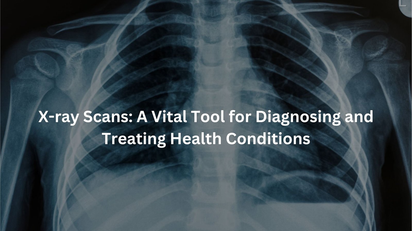
In cancer care, X-rays play a key role. They can help detect tumours, monitor the progress of cancer treatments, and check if cancer has spread. A regular chest X-ray might show signs of lung cancer, while a mammogram (an X-ray of the breast) is a key part of detecting breast cancer.
Pulmonology:
For lung conditions, X-rays are invaluable. They can be used to diagnose pneumonia, lung infections, and even lung cancer. X-rays allow doctors to see the size and shape of the lungs and detect any abnormal growths or infections.
Radiation Safety in X-ray Scans
Credits: Medical Centric
While X-rays are an important tool for doctors, they do come with a risk—radiation exposure. However, the amount of radiation used in medical X-rays is very small and controlled to minimise risk. That said, it’s important to always weigh the benefits of the scan against the risks.
Types of Radiation:
X-rays are considered ionising radiation, which means they can remove electrons from atoms, potentially leading to cellular damage. However, the risk from a single X-ray scan is generally low, and the procedure is extremely beneficial for diagnosing health issues early.
Safety Measures:
- Lead aprons: Often, patients are asked to wear lead aprons to protect sensitive areas of their body from unnecessary radiation exposure.
- Minimising exposure: Healthcare professionals only perform X-ray scans when absolutely necessary, and they’ll use the lowest possible radiation dose to achieve accurate results.
- Informed consent: Patients are always informed about the potential risks of radiation before the procedure.
Although there is a small lifetime risk of developing cancer from repeated exposure, the safety measures in place significantly reduce this risk.
Bone Density and X-rays
One of the most common uses of X-rays is in measuring bone density, particularly through a DEXA scan. This type of X-ray is used to evaluate the strength of bones and detect conditions like osteoporosis. Osteoporosis weakens bones and makes them more likely to break, and a bone density test can help doctors assess the condition early on.
As people age, their bones naturally lose mass, so measuring bone density is particularly important in older adults. It’s a simple and effective way to catch bone-related health issues before they become serious. The risks of these scans are minimal, but the benefits are significant, especially for people at high risk of osteoporosis.
X-rays in Cancer Diagnosis and Treatment
X-rays are crucial in both the diagnosis and treatment of cancer. They’re used to detect the presence of tumours, track cancer progression, and even assist in radiation therapy.
Detecting Tumours:
X-rays can be used to detect abnormalities in organs, such as the lungs or breasts. For example, a chest X-ray can reveal early signs of lung cancer, while a mammogram helps spot breast cancer.
Monitoring Cancer Progression:
After the diagnosis of cancer, X-rays are often used to monitor how well the treatment is working. If a tumour is shrinking, it will show up differently on the X-ray image.
Radiation Therapy:
In cancer treatment, X-rays are used in radiation therapy. The high doses of radiation kill or shrink cancer cells by damaging their DNA, preventing them from growing and dividing.
Pediatric X-ray Considerations
Children are more sensitive to radiation, so extra care is needed when performing X-ray scans. Pediatric X-rays are done with the lowest possible dose of radiation, and healthcare providers take special precautions to ensure the child’s safety.
For example, healthcare professionals may use lead shields to protect the child’s reproductive organs or other sensitive areas. Additionally, pediatric X-ray scans are often done with special equipment to ensure the images are clear while keeping the radiation dose as low as possible.
Innovations and Future of X-ray Technology
The field of X-ray technology is constantly advancing. Modern X-ray machines are getting better at providing high-quality images with less radiation. New detectors allow for clearer images, and improved computer software helps doctors interpret these images with more precision.
Artificial Intelligence:
AI is also starting to play a bigger role in interpreting X-ray scans. AI can help radiologists quickly identify patterns that may be missed by the human eye, improving accuracy and efficiency.
Future Trends:
The future of X-ray technology looks promising. There’s potential for even lower radiation doses, better image quality, and faster scanning techniques. With these advancements, X-rays will continue to be a key part of diagnosing and treating a wide variety of health conditions.
Conclusion
As X-ray technology continues to improve, its role in diagnosing and treating conditions will only grow. Understanding the different types of X-ray scans, the safety measures in place, and their wide-ranging applications can help individuals make informed decisions about their health.
Whether it’s detecting a broken bone, identifying a tumour, or assessing bone density, X-ray scans are an essential part of healthcare that continue to evolve to meet patient needs.
FAQ
What are X-ray scans used for?
X-ray scans are used in healthcare to help doctors see inside the body and examine bones and internal structures. They use a special type of radiation to create images that can reveal problems like bone fractures or infections.
How do X-ray scans work?
X-ray scans work by passing a form of radiation through the body. The X-ray machine sends out a beam of radiation, and the X-ray detectors on the other side capture how the radiation passes through. Different parts of the body, like bone and soft tissue, absorb the radiation in different amounts, creating an image for doctors to analyse.
Are X-ray scans safe?
Yes, X-ray scans are generally safe. They use a very low level of radiation, which is why healthcare providers make sure to only use them when necessary. The amount of radiation you receive is very small and the risk of harm is low, especially when the scans are used correctly.
Can X-rays detect soft tissue injuries?
X-rays are better at detecting problems with bones and bony structures. Soft tissue like muscles, fat, and organs might not show up clearly on a standard X-ray, which is why other types of medical imaging like Magnetic Resonance Imaging (MRI) may be used for soft tissue injuries.
Can X-rays see through metal objects?
X-rays can sometimes have difficulty seeing through dense materials, like metal objects. If there are metal objects in the body, such as a metal implant, it can block the X-ray from creating a clear image of the surrounding areas. However, doctors can adjust the X-ray machine to get the best possible view.
How does the X-ray machine produce images?
The X-ray machine uses a special X-ray source that sends a controlled radiation beam through the body. The radiation passes through the body and is detected by X-ray detectors on the other side. The X-ray images created are then analysed by healthcare providers to check for any issues.
What is the difference between a chest X-ray and a bone scan?
A chest X-ray focuses on the organs and bones in the chest area, like the heart and lungs, and is typically used to check for conditions like pneumonia or lung diseases. A bone scan, on the other hand, is used to look for issues with bones, such as infections or fractures.
Can X-ray scans be used for paediatric patients?
Yes, X-ray scans can be used for paediatric patients, but healthcare providers take extra care to minimise radiation exposure, especially for children. Paediatric patients may need fewer X-rays than adults, and they might use the lowest doses possible to get clear images.
Do X-ray scans show all body parts?
X-ray scans can show bones and some internal structures, but they may not provide clear images of soft tissue, organs, or areas that contain dense material, like metal implants. For these types of images, other medical imaging techniques might be used.
Are there any risks associated with radiation exposure from X-ray scans?
While X-ray scans do involve exposure to a type of radiation, the doses are very low. For most people, the benefits of having an X-ray scan far outweigh the risks. However, healthcare providers try to use the lowest doses of radiation necessary to get the needed image, and only perform X-ray exams when they are truly needed.
Can X-ray technology be used to check for bone fractures?
Yes, X-ray scans are commonly used to detect bone fractures. The radiation in the X-ray machine can easily pass through soft tissues but is absorbed by bones, allowing doctors to see fractures, breaks, or other issues with bone density.
What types of radiation are involved in X-ray scans?
X-ray scans use a type of radiation called ionising radiation. This type of radiation has enough energy to remove electrons from atoms, which can sometimes cause damage to cells. However, the level of radiation used in X-ray scans is very small and the risk of harm is low when the scans are used properly.
How do doctors know how much radiation to use?
Doctors and healthcare providers are trained to ensure that the amount of radiation used in X-ray scans is the minimum needed to get clear and accurate images. This helps minimise the amount of radiation exposure while still providing valuable information for diagnosis.
Can X-rays be used for bone density tests?
Yes, dual-energy X-ray absorptiometry (DXA) is a type of X-ray scan that is commonly used for bone density tests. This type of test helps doctors assess bone mineral density and check for conditions like osteoporosis.
References
- https://www.betterhealth.vic.gov.au/health/conditionsandtreatments/x-ray-examinations
- https://www.health.gov.au/topics/diagnostic-imaging/about


