

X-rays let doctors see inside our bodies, helping spot broken bones or hidden issues. Learn how this safe, fascinating energy helps diagnose health problems!
X-rays are special energy waves that let doctors look inside our bodies. They can show if bones are broken or if there’s a problem inside that we can’t see. When doctors use X-rays, they get a clear picture of what’s going on, making it easier to diagnose health issues.
It’s pretty amazing how these invisible beams can help us stay healthy. Plus, X-rays are safe when done by professionals. If you’re curious about how X-rays work or their importance in medicine, keep reading to discover more about this vital tool in healthcare!
Key Takeaway
- X-rays help doctors see inside the body to find problems.
- There are different types of X-rays for different needs, like chest X-rays and dental X-rays.
- Preparing for an X-ray is easy, and the process is quick and safe.
X-ray Scans
When you visit the doctor, they might need to look inside your body, and that’s where X-rays come in. An X-ray is like a photo, but instead of light, it uses invisible beams to create an image of your bones and organs. It’s quick and doesn’t hurt.
Here’s how it works: the X-ray machine sends beams through your body. Soft tissues, like muscles and organs, let most of the beams pass through, so they appear grey on the image. Bones, which block more beams, show up white. This contrast helps doctors spot things like broken bones or unusual objects inside you.
X-rays are super useful in emergencies, like after a fall or accident, because they can show injuries fast. They’ve been around for over 100 years, so they’re a trusted tool. If you ever need one, just stay still—it’ll be done in seconds and might help your doctor treat you better.
Diagnostic X-rays
When someone has pain, like in their leg or arm, a doctor might ask for a diagnostic X-ray. It’s a quick and simple way to see inside the body without surgery. X-rays are invisible energy beams that pass through the body to create an image. Bones block the X-rays, so they show up white, while softer tissues like muscles and organs appear grey.
Here’s how it works:
- The X-ray machine sends beams through your body.
- Bones block the beams and show as white.
- Softer tissues let the beams through, appearing grey.
Doctors use X-rays to check for fractures or other issues, like foreign objects. It’s fast and doesn’t hurt. A friend of mine had one after a fall, and it only took a few minutes to confirm nothing was broken. If your doctor suggests an X-ray, it’s nothing to stress about. It’s just a safe way to get answers quickly.
How X-rays Work
X-rays are pretty fascinating, aren’t they? They use invisible beams of X-ray photons (a type of radiation) to peek inside the body. These photons pass through tissues, creating an image that shows what’s hidden underneath the skin. It’s like magic, but science.
Here’s how it works:
- The X-ray photons hit your body.
- Dense stuff like bones blocks most of the X-rays, so they show up white on the image.
- Softer tissues, like muscles or organs, let more X-rays pass through, appearing in shades of grey.
The X-ray machine (called an X-ray tube) makes the beams and captures the image in seconds. My mate Sarah broke her arm playing footy, and her X-ray showed the fracture straight away—a clear white line where the bone snapped. It’s quick, painless, and gives doctors the info they need. If you ever need one, it’ll be over before you know it.
X-ray Uses

X-rays are a big deal in healthcare. They’ve made it easier to figure out what’s wrong inside the body without needing surgery. I remember when my cousin fell off his bike and thought he’d broken his leg. The doctor used an X-ray, and in just a few minutes, they knew it was just a bruise, not a fracture. Amazing, right?
Here’s what X-rays are good for(1):
- Bones: They show fractures or breaks clearly.
- Soft tissues: While not as clear as bones, they can still catch issues in muscles or organs.
- Lungs: Chest X-rays help spot infections or even cancer.
- Teeth: Dental X-rays check for cavities or gum problems.
- Foreign objects: If you swallow something strange, X-rays can find it fast.
This tech saves time and lives. It’s quick, painless, and gives doctors the answers they need without cutting you open. Pretty brilliant, I reckon.
X-ray Images
X-ray images are like windows into the body. They let doctors see inside without needing to cut you open. I remember when my mate sprained her ankle during netball. The doctor ordered an X-ray, and within minutes, we could see her bones on the screen—bright white against shades of grey. Thankfully, no breaks, just a sprain.
X-rays are used for heaps of things:
- Bones: Spotting fractures, breaks, or infections.
- Soft tissues: Showing enlarged organs or fluid in the lungs (though not as sharp as bones).
- Treatment checks: Seeing how well injuries or illnesses, like pneumonia, are healing.
- Foreign objects: Finding things like swallowed coins or metal bits.
The process is quick and painless. You stand still, maybe hold your breath, and that’s it. If you ever need one, don’t stress—it’s just a fast way for doctors to figure out what’s going on inside.
Chest X-rays
When someone’s lungs don’t feel right—maybe from coughing or trouble breathing—a doctor might suggest a chest X-ray(2). It’s quick and shows what’s happening inside. I remember when a friend of mine, feeling sick for weeks, got one. The X-ray revealed early pneumonia. That image helped the doctors act fast.
Chest X-rays are handy for spotting problems like:
- Lung issues (fluid, infections, or even tumours).
- Heart problems (like an enlarged heart or fluid buildup).
- Broken ribs or chest bone issues.
- Breathing troubles (pneumonia or a collapsed lung).
The process is easy. You stand still, take a deep breath, and hold it. The machine snaps a picture, and within minutes, the doctor has a clear view. It’s not scary, just helpful. If you’re ever feeling unsure about your chest or breathing, don’t wait. That simple X-ray might be the key to figuring out what’s wrong.
Dental X-rays
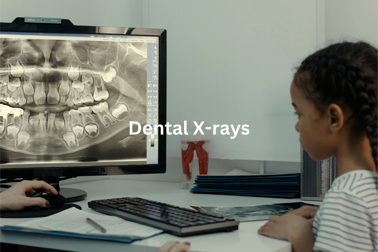
Anyone who’s been to the dentist knows that an X-ray is probably part of the visit. I remember my first one—biting down on that hard plastic piece while the machine hummed softly. It felt strange but wasn’t painful. Those X-rays, though, they’re like a map of your teeth.
Dentists use X-rays for more than just spotting cavities (though they’re great for that). They can also:
- Detect gum disease by showing bone changes.
- Check tooth roots for infections or damage.
- Look at your jawbone for hidden problems.
- Find wisdom teeth that might be stuck under the gums.
There are two main types: bitewing X-rays (you bite on a tab) and panoramic X-rays (a full-mouth view). Both are quick, painless, and done in minutes. So, if your dentist suggests an X-ray, it’s worth it. They help catch issues early, saving you trouble later. A few minutes now can mean healthier teeth for years.
X-ray Radiation
Sources: University of South Australia.
It’s normal to feel a bit uneasy when you hear the word “X-ray.” Radiation can sound scary, right? But the truth is, the radiation from medical X-rays is very small. Actually, it’s less than what we’re exposed to daily just by being outside.
X-rays are a type of electromagnetic radiation (like light, but stronger). They help doctors see inside the body, which is super useful. But because too much radiation can be harmful, X-rays are only used when needed. Plus, there are lots of safety measures.
Here’s how it works:
- Low dose: Medical X-rays use minimal radiation.
- Protection: Lead aprons and careful aiming keep exposure low.
- Quick: The process takes just seconds.
- Everyday exposure: One X-ray equals less radiation than daily life (sunlight, soil, etc.).
So, whether it’s for a broken arm or a dental check-up, X-rays are safe and helpful when used properly.
Preparing for X-rays
Getting ready for an X-ray isn’t complicated. It’s more like ticking off a short checklist. I’ve had one before (for a sprained ankle), and honestly, it’s over before you know it.
First, you’ll probably need to change into a hospital gown. Anything metal—like jewellery or belts—needs to come off since it can interfere with the image. If your X-ray involves contrast material (used for clearer images of certain areas like the stomach), you might be asked to fast for a few hours beforehand. No food, no drinks.
And if there’s even a chance you’re pregnant, let the technician know. They might recommend a different test, like an MRI, to avoid radiation exposure. It’s quick and straightforward. My last X-ray took about 10 minutes, tops. Just be upfront about your health history, and you’ll be fine. Relax—it’s just a snapshot of what’s happening inside.
Interpreting X-rays
Looking at an X-ray is like solving a puzzle. Each shade of grey tells a story about what’s happening inside your body. Doctors are trained to read these images, spotting things like fractures, infections, or even tumours. It’s not just random shadows—it’s a map of your health.
When reading an X-ray, doctors focus on details(3):
- Fractures: Breaks in the bone stand out clearly.
- Tumours: Unusual shapes or masses might suggest growths.
- Infections: These can appear as areas of strange density or shadowing.
Sometimes, they’ll compare your X-ray to an older one to see if anything’s changed. If they spot something unusual, they might order more tests, like a CT scan, or discuss treatment options with you. It’s not magic, just science. But it’s pretty amazing how a simple image can help doctors figure out what’s wrong and how to help you feel better.
FAQ
What are X-ray beams and how do they differ from other forms of radiation?
X-ray beams are a type of electromagnetic wave, similar to visible light but with a much shorter wavelength. Unlike visible light, X-ray beams are a form of radiation that can penetrate the human body, making them useful for medical imaging procedures like X-ray exams and X-ray crystallography.
How are radiation doses and levels of radiation exposure measured during X-ray procedures?
The amounts of radiation and doses of radiation used in X-ray imaging are carefully controlled. Medical professionals measure background radiation, radiation doses, and exposure to radiation to ensure patients receive the lowest effective dose needed for diagnostic purposes. This helps minimise the risk of adverse biological effects like cancer.
What precautions should pregnant patients or those with metal objects in their body take during X-ray procedures?
Pregnant patients may need to take extra precautions during X-ray exams to protect the fetus from radiation exposure. Patients with metal objects in their body, like dental fillings or joint replacements, should inform their healthcare provider, as these can affect the quality of X-ray images.
How do X-ray imaging devices create detailed cross-sectional images of the human body?
X-ray imaging devices, such as X-ray detectors and X-ray microscopes, use X-ray emission to capture images of the body’s internal structures. The amount of X-ray beams absorbed by different tissues creates shades of grey in the resulting X-ray photograph, allowing medical professionals to identify bones, organs, and other features.
What are the benefits and risks of X-ray procedures compared to other medical imaging techniques like magnetic resonance imaging (MRI)?
X-rays are a valuable diagnostic tool, allowing doctors to see detailed images of the body’s internal structures. However, X-rays do expose patients to a small amount of ionising radiation, which can increase the cumulative risk of developing conditions like bone cancer or lung cancer over time. In contrast, MRI uses magnetic fields and radio waves, avoiding the use of radiation.
Conclusion
You know, X-rays are really cool tools that help doctors look inside our bodies. They’re safe and quick, which is super important when figuring out health problems. Like, from chest X-rays to dental X-rays, they’re used in many ways. X-ray technology keeps getting better too, making it easier for doctors to see what’s going on. So, when you hear about X-rays, think about how they help keep us healthy in so many ways!
References
- https://www.healthdirect.gov.au/x-rays
- https://www.svhlunghealth.com.au/procedures/imaging/chest-x-ray
- https://researchoutput.csu.edu.au/en/publications/chest-x-ray-image-interpretation


