A cardiac nuclear scan — most commonly a myocardial perfusion scan — uses a small radioactive tracer to show how well blood reaches your heart muscle, usually comparing the heart at rest and under 'stress' (exercise or medication). It helps assess chest pain and coronary artery disease, and check the heart after a heart attack. The imaging is painless; the stress part is fully supervised by medical staff.
Key takeaways
- A cardiac nuclear scan shows blood flow to the heart muscle.
- It usually compares the heart at rest and under stress.
- It helps assess chest pain and coronary artery disease.
- The stress part is supervised by medical staff throughout.
Cardiac nuclear imaging is a way of seeing not just what the heart looks like, but how well blood is reaching the heart muscle — a key question when chest pain or heart disease is being investigated.
What it shows
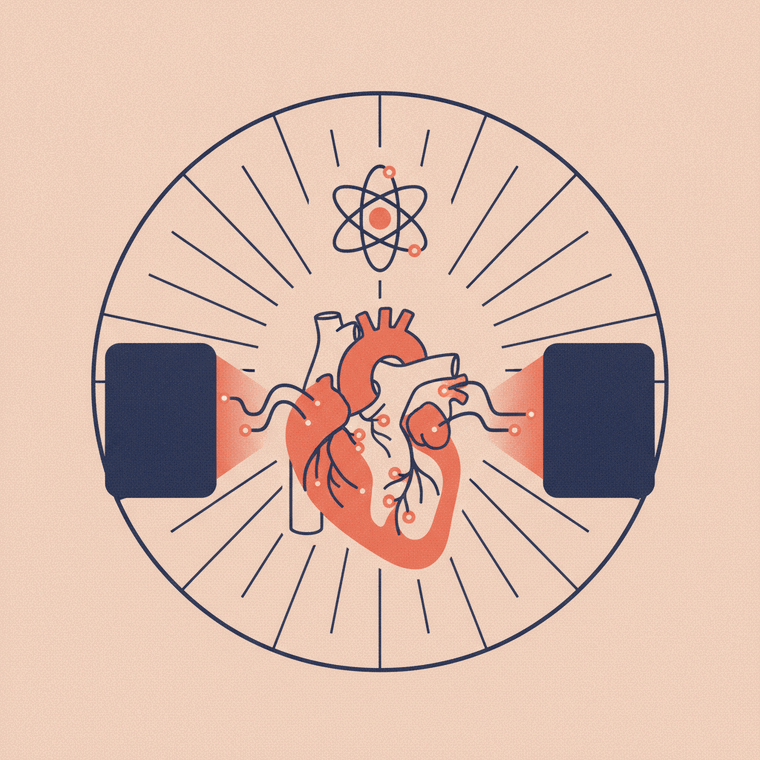
The most common test is a myocardial perfusion scan. A small radioactive tracer is given, and a gamma camera shows how well it — and therefore blood — is reaching different parts of the heart muscle.[1] Areas that don’t receive enough blood show up as having reduced uptake.
It’s used to:[1]
- Assess chest pain and investigate coronary artery disease
- Measure the extent of a narrowing in the heart’s arteries
- Check how the heart is coping after a heart attack or a procedure (such as bypass surgery or a stent)
A related test, MUGA, checks the heart’s pumping function — for example before and after chemotherapy.[1]
Rest and stress
A perfusion scan usually compares two sets of images:[1]
- at rest, and
- under stress — either physical exercise (on a treadmill or bike) or, if you can’t exercise, a medication that mimics its effect on the heart.
Comparing the two shows whether parts of the heart muscle are short of blood only when working hard — a sign of narrowed coronary arteries that may be missed at rest.
What to expect
The tracer is injected, and the camera takes pictures of your chest. The stress portion is carried out with medical staff and ECG monitoring throughout, so any symptoms are managed promptly.[1] The dose is in the usual nuclear medicine range. There may be specific preparation — such as avoiding caffeine beforehand, or adjusting some medications — so follow your clinic’s instructions.
Frequently asked questions
What is the ‘stress’ part of the scan?
It’s a way of making your heart work harder — by exercise or a medication — so the scan can reveal areas short of blood flow that look normal at rest. It’s fully supervised with heart monitoring.[1]
Is a cardiac nuclear scan safe?
Yes — it uses a small radiation dose, and the stress portion is monitored by staff throughout. Tell the team if you could be pregnant or are breastfeeding.[1]
How is this different from an echocardiogram?
An echocardiogram is an ultrasound of the heart (no radiation) showing structure and pumping. A cardiac nuclear scan shows blood flow to the heart muscle using a tracer. They answer different questions and are sometimes used together.[1]
Sources
- RadiologyInfo.org (RSNA & ACR) — General Nuclear Medicine (heart) — www.radiologyinfo.org/en/info/gennuclear
- RANZCR / InsideRadiology — Nuclear medicine (Cain) — www.insideradiology.com.au/nuclear-medicine/