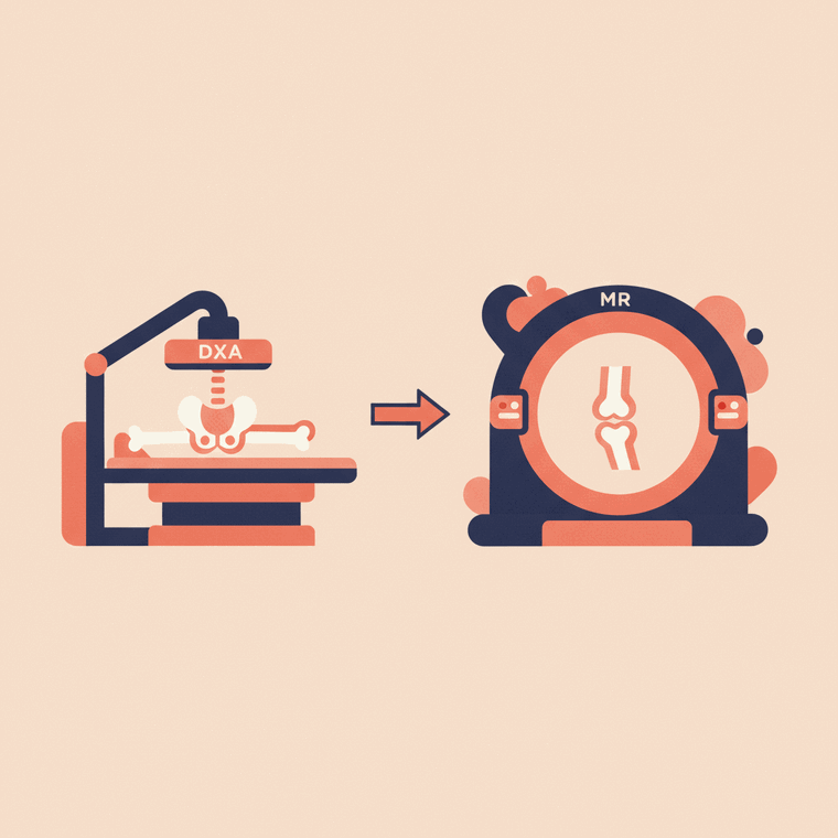
A DXA scan measures bone density — how strong your bones are — to diagnose and monitor osteoporosis. An MRI gives detailed pictures of soft tissues and structures (like bone marrow, joints, discs and nerves). They answer different questions, so you'd have a DXA for bone strength and an MRI for detailed structural problems. They're not interchangeable.
Key takeaways
- DXA measures bone density (strength) — for osteoporosis.
- MRI shows detailed soft tissue and structure, with no radiation.
- They answer different questions and aren't interchangeable.
- DXA is quick and very low-dose; MRI is longer and radiation-free.
A DXA scan and an MRI are sometimes confused, but they do completely different jobs — so it’s not really a case of one being “better.”
DXA measures bone strength
A DXA scan measures bone mineral density — how strong (or thin) your bones are. It’s the gold-standard test for diagnosing and monitoring osteoporosis, and it’s quick, painless and very low-dose.[1] It gives a number (your T-score), not a detailed picture.
MRI shows detailed structure
An MRI produces detailed images of soft tissues and structures — bone marrow, joints, cartilage, discs, nerves and more — using magnets and radio waves, with no radiation.[3] It shows what something looks like in fine detail, rather than measuring density.
When you’d have each
- You’d have a DXA to find out whether you have osteoporosis or to monitor your bone density over time.
- You’d have an MRI to investigate a specific structural problem — a joint injury, a disc, a suspicious area in the bone, or marrow disease.
They can even be complementary — but they’re not interchangeable. (For a spine with existing fractures or arthritis, where DXA is less reliable, a CT-based method may occasionally be used instead.)[1]
Frequently asked questions
Can an MRI measure bone density instead of a DXA?
Not in the standard way DXA does. DXA is the established test for measuring bone density and diagnosing osteoporosis; MRI shows structure, not a density score.[1]
Which has more radiation, DXA or MRI?
DXA uses a very low dose of radiation; MRI uses none. But they’re used for different purposes, so the choice is about what needs to be assessed.[3]
Sources
- RadiologyInfo.org (RSNA & ACR) — Bone Density Scan (DEXA/DXA) — www.radiologyinfo.org/en/info/dexa
- RANZCR / InsideRadiology — Bone mineral density scan (DXA) (Hendrich) — www.insideradiology.com.au/bone-mineral-density-scan-hp/
- RANZCR / InsideRadiology — Magnetic resonance imaging (MRI) — www.insideradiology.com.au/mri-hp/