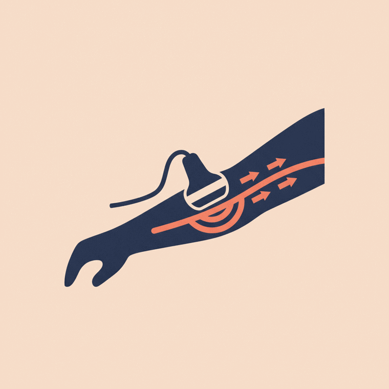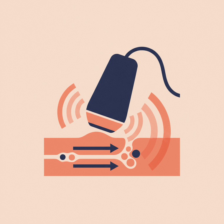
A pelvic ultrasound checks the organs in the pelvis — in women the uterus and ovaries, in men the bladder and prostate. It uses no radiation (sound waves) and is done either over the lower tummy (transabdominal, needing a full bladder) or, for a closer view, internally (transvaginal in women). It's painless and used for pelvic pain, abnormal bleeding, cysts, fibroids and bladder problems.
Key takeaways
- A pelvic ultrasound checks the uterus and ovaries, or the bladder and prostate.
- It uses no radiation — sound waves.
- The abdominal approach needs a full bladder; an internal scan gives a closer view.
- It's used for pelvic pain, bleeding, cysts, fibroids and bladder issues.
A pelvic ultrasound is a common, safe scan of the organs low in the abdomen. What it looks at depends on why you’re having it.
What it shows
A pelvic ultrasound uses sound waves — no radiation — to check the pelvic organs:[1]
- in women: the uterus (and its lining), the ovaries, and the causes of pelvic pain, abnormal bleeding, cysts or fibroids;
- in men: the bladder and prostate, and how well the bladder empties;
- in anyone: the bladder, and sometimes to guide a procedure.
The two ways it’s done
There are two approaches, often used together:[2]
- Transabdominal — the probe moves over your lower tummy with gel. This needs a full bladder, which lifts the bowel out of the way and gives a clear window onto the organs. You’ll be asked to drink water beforehand and not go to the toilet.
- Transvaginal (women) — a slim internal probe gives a closer, clearer view; for this you empty your bladder first. It’s your choice — see our guide to the transvaginal ultrasound.
What to expect
It’s painless and takes about 20–30 minutes. For the abdominal part, gel is applied and the probe is moved over your lower abdomen. There’s no radiation and no aftercare — you can go straight back to normal activities.[1] For preparation details, see ultrasound preparation.
Frequently asked questions
Why do I need a full bladder for a pelvic ultrasound?
For the abdominal approach, a full bladder pushes the bowel aside and creates a clear “window” onto the uterus, ovaries or prostate. (An internal transvaginal scan is the opposite — you empty your bladder first.)[1]
Is a pelvic ultrasound the same as a transvaginal one?
A transvaginal ultrasound is one type of pelvic ultrasound — the internal approach that gives a closer view. A pelvic ultrasound may be done over the abdomen, internally, or both.[2]
Does a pelvic ultrasound use radiation?
No — it uses sound waves, so there’s no radiation, which is one reason it’s safe in pregnancy.[1]
Sources
- RadiologyInfo.org (RSNA & ACR) — General Ultrasound — www.radiologyinfo.org/en/info/genus
- RANZCR / InsideRadiology — Ultrasound (Coombs) — www.insideradiology.com.au/ultrasound-hp/
- healthdirect (Australia) — Ultrasound — www.healthdirect.gov.au/ultrasound